
What you need to know
- 5.5chloroplastThe organelle where photosynthesis takes place. It contains thylakoids and stroma. structure in relation to the role in photosynthesisThe process in which plants use light energy to make glucose from carbon dioxide and water, releasing oxygen..
- 5.6absorption spectrumA graph of how much light a pigment absorbs at each wavelength. and action spectrumA graph of the rate of photosynthesis at each wavelength of light..
- 5.7Separate chloroplast pigments by chromatographyA method that separates the substances in a mixture by how far each moves with a solvent. and identify them using Rf valueDistance moved by a substance ÷ distance moved by the solvent front, in chromatography..
4A 2 asked you to recognise a chloroplast. This lesson is about why the parts are there. Cyclic and non-cyclic photophosphorylation remain 5A 3. Core Practical 10 measures rate, not Rf.
By the end, you should be able to…
- Link envelopeAn outer covering membrane. The nucleus and chloroplasts have a double-membrane envelope; some viruses, such as HIV, have a lipid envelope taken from the host cell., stromaThe fluid inside a chloroplast, where the light-independent reactions take place., thylakoidA flattened membrane sac in a chloroplast, where the light-dependent reactions take place. and granumA stack of thylakoids in a chloroplast. to the two stages of photosynthesis.
- Distinguish an absorption spectrum from an action spectrum.
- Calculate Rf and explain why more than one pigment appears.
Build your understanding
A chloroplast is a partitioned workshop
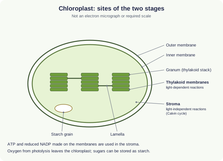
A chloroplastThe organelle where photosynthesis takes place. It contains thylakoids and stroma. has a double envelopeAn outer covering membrane. The nucleus and chloroplasts have a double-membrane envelope; some viruses, such as HIV, have a lipid envelope taken from the host cell.. Inside, the stromaThe fluid inside a chloroplast, where the light-independent reactions take place. is the fluid containing Calvin-cycle enzymeA globular protein that acts as a biological catalyst, speeding up a specific reaction., chloroplast DNADeoxyribonucleic acid: a double-stranded polynucleotide that carries the genetic code., ribosomeA small organelle made of rRNA and protein, where translation takes place. and often starchThe storage polysaccharide in plants, made of amylose and amylopectin. It is compact and insoluble, so it does not affect water potential. grains. thylakoidA flattened membrane sac in a chloroplast, where the light-dependent reactions take place. are flattened membrane sacs. Stacks of thylakoids are granumA stack of thylakoids in a chloroplast.; membranes joining grana are lamellae. Photosynthetic pigments and electron-transport proteins sit in the thylakoid membranes.
A chloroplast is a partitioned workshop (continued)
light-dependent reactionsThe reactions on the thylakoid membranes that use light energy to make ATP and reduced NADP, splitting water and releasing O₂. run on those membranes: large surface areaThe total area of a surface. A larger surface area gives a faster rate of diffusion., short diffusion distanceThe distance molecules have to travel. A shorter distance gives a faster rate of diffusion. into the thylakoid space, pigments held next to electron carriers. light-independent reactionsThe reactions in the stroma (the Calvin cycle) that use ATP and reduced NADP to fix CO₂ and make GALP. run in the stroma, where carbon dioxide, ATPAdenosine triphosphate: the molecule that releases energy for cell processes when it is hydrolysed to ADP and Pi. and reduced NADPNADP that has accepted hydrogen in the light-dependent reactions. It supplies the hydrogen to reduce GP to GALP. can meet RUBISCOThe enzyme that catalyses carbon fixation, joining CO₂ to RuBP. and the other enzymes. Structure is only useful in an answer if you finish with that role.
Absorption spectrum and action spectrum
An absorption spectrumA graph of how much light a pigment absorbs at each wavelength. is a graph of how much light a pigment, or a mixture of pigments, absorbs at each wavelength. Chlorophylls absorb strongly in blue-violet and red, and poorly in green, which is why many leaves look green. Accessory pigments, including carotenoids, absorb other wavelengths and can pass energy towards chlorophyllThe green pigment in chloroplasts. It absorbs mainly red and blue light. a.
Absorption spectrum and action spectrum (continued)
An action spectrumA graph of the rate of photosynthesis at each wavelength of light. is a graph of the rate of photosynthesisThe process in which plants use light energy to make glucose from carbon dioxide and water, releasing oxygen. against wavelength. It is similar to the combined absorption spectrum because absorbed photons can drive the process, but it is not a photocopy. Some absorbed wavelengths are less efficiently used, and a leaf contains more than one pigment. Do not define one graph with the name of the other. Some books quote photosystem II near 680 nm and photosystem I near 700 nm. Those peaks are a reason two photosystemA cluster of chlorophyll and other pigments with proteins in the thylakoid membrane that absorbs light and passes energy to a reaction centre. together use more of the red end. They are enrichment, not numbers 5.6 requires you to memorise.
Chromatography and Rf
Chloroplast pigments can be separated because they differ in solubility in the solventA liquid in which other substances (solutes) dissolve. Water is a good solvent for ions and polar molecules. and in how strongly they adsorb to the paper or thin layer. A concentrated extract is spotted on a pencil origin, not ink. The solvent is allowed to rise, then the solvent front is marked before it dries. Follow the school’s solvent and fume-hood rules; this is knowledge of a method, not a licence to mix solvents unsupervised.
Rf = distance moved by the pigment ÷ distance moved by the solvent, both measured from the origin to the spot centre and the front. Rf has no unit and is between 0 and 1. It is comparable only in the same solvent system. Typical school chromatograms show chlorophyll a, chlorophyll b and yellow carotenoids; do not memorise a universal Rf table, because changing the solvent changes the values.
Follow the process
Pencil origin → spot a concentrated pigment extract and let it dry → stand the paper so the solvent is below the origin → the solvent rises and the pigments separate → mark the solvent front before it dries → measure both distances from the origin → Rf = pigment distance ÷ solvent distance.
These pigments separate because they differ in solubility in the solvent and in how strongly they adsorb to the paper. Do not explain this chromatogram as a separation by charge. Charge is the idea behind gel electrophoresisSeparating DNA fragments by size: they move through a gel towards the positive electrode, and smaller fragments move further., which is a different method.
Diagram B
NOT: this chromatogram is not separated by charge. Charge is the idea behind gel electrophoresis.
- Measure to the centre of the spot and to the solvent front
- Rf is comparable only in the same solvent
- School plates often show chlorophyll a, chlorophyll b and carotenoids
- Do not memorise a universal Rf table
- Changing the solvent changes the Rf values
Two traps this diagram blocks
- Do not use an ink origin or start the solvent above the spot.
- Do not quote one Rf as true for every solvent.
From the diagram alone
Question
A pigment spot travels 24 mm and the solvent front travels 60 mm from the same origin. Calculate Rf and state one reason a published Rf may not match yours. [3]
Show a model answer
Rf = 24/60 = 0.40, with no unit. A different solvent, or a different paper, changes how far the pigment moves, so published values are not universal.
| Graph | What is plotted | What it tells you |
|---|---|---|
| Absorption spectrum | Absorbance against wavelength | Which wavelengths a pigment takes up |
| Action spectrum | Rate of photosynthesis against wavelength | Which wavelengths actually drive the process |
| Why they differ | A leaf is a mixture; not all absorbed energy is used equally | Similarity supports pigment involvement; identity is not required |
Make your reasoning visible
Build the explanation
X happens because Y, leading to Z. X: the action spectrumA graph of the rate of photosynthesis at each wavelength of light. is high in blue-violet and red and low in green. Y: those are the wavelengths chlorophylls absorb, so they can excite electrons. Z: photosynthesisThe process in which plants use light energy to make glucose from carbon dioxide and water, releasing oxygen. is fast where absorption is strong and slow where light is mostly reflected. Incomplete: “The action spectrum is the absorption spectrumA graph of how much light a pigment absorbs at each wavelength..” Improved: “The action spectrum is rate against wavelength. It resembles the absorption spectrum because only absorbed light can drive the reactions, but the two graphs measure different things.”
Finish structure with function
“granumA stack of thylakoids in a chloroplast. are stacks” scores little until you say they hold a large membrane area of pigments and carriers.
Measure from the origin
Rf uses the same starting line for pigment and solventA liquid in which other substances (solutes) dissolve. Water is a good solvent for ions and polar molecules.. Do not measure from the bottom of the paper.
Green is a clue
Poor absorption of green light is why the action spectrum is also low there, not because green light “does not exist” in sunlight.
Key explanations
- The origin is in pencil. Ink would run and confuse the pigments.
- Mark the solvent front before it dries, or the denominator cannot be measured.
- Do not explain this separation as movement by electrical charge.
Avoid this misconception
The claim: A leaf contains only chlorophyllThe green pigment in chloroplasts. It absorbs mainly red and blue light., and the absorption spectrum is the action spectrum.
The correction: Several pigments are present and can be separated. The two spectra are related but defined differently.
Why are leaves green, when green light is the colour they use least?
Chloroplasts hold several pigments in their thylakoid membranes. Each absorbs some wavelengths strongly and others hardly at all.
What the model shows
A chloroplast lit by one wavelength at a time, with the absorption and action spectra. Then paper chromatography that separates the pigments, with Rf measured from the origin.
What to look for
Which colours the thylakoids absorb, and how the rate of photosynthesis follows. How far each spot moves compared with the solvent front.
What it tells us
Chlorophylls absorb blue-violet and red light and reflect green. The action spectrum resembles the absorption spectrum. A leaf has more than one pigment, and Rf = pigment distance ÷ solvent distance (4.2 ÷ 7.0 = 0.60).
Be careful what you claim
Illustrative curves and fictional solvents. Rf values only compare within one solvent system.
Try it in the model: drag the wavelength slider from blue to green to red and watch the thylakoids.
Chloroplast pigments: spectra and chromatography
Shine one wavelength at a time on a chloroplast and compare the absorption and action spectra, then separate a leaf extract by chromatography and calculate Rf. Curves and solvents are illustrative.
What you see
- Spectra: a chloroplast with its double envelope, stroma, grana, thylakoids and lamellae in a beam of light of the wavelength you choose. The graph shows the absorption spectra and, if you turn it on, the action spectrum.
- Chromatography: a strip of paper in a tank, the solvent rising past a leaf-extract spot, a ruler from the origin and an Rf chart.
How to use it
- Under Part, choose Spectra or Chromatography.
- Spectra: drag the wavelength slider, choose which pigments to plot, and turn the action spectrum on or off.
- Chromatography: choose the origin, the solvent level and the solvent, run it, remove the paper and mark the front, then choose a spot to measure.
What it shows
- How chloroplast structure suits the two stages of photosynthesis (5.5).
- The difference between an absorption spectrum and an action spectrum (5.6).
- Separating chloroplast pigments by chromatography and identifying them by Rf (5.7).
Limits of the model
- The spectra are smooth illustrative curves, not measured data. Real peaks depend on the solvent and the leaf.
- Solvents X and Y and their Rf values are fictional. Follow your school’s solvent and fume-cupboard rules in a real practical.
- The chloroplast is a schematic, not an electron micrograph.
Guided tour · step 1 of 13
- In Light, just below these steps, set Wavelength (400–700 nm) between 420 and 480 nm.
- Watch the thylakoids and the card in the simulator.
Chlorophylls and carotenoids absorb blue-violet light strongly. The pigments sit in the thylakoid membranes, where the light-dependent reactions happen.
Light
Questions
Check your understanding
Try each question before opening the suggested answer.
Practice question
Retrieval and explanation · suggested answer
Explain how thylakoid stacking supports the light-dependent reactions.
Show suggested answer
Grana provide a large membrane area for pigments, electron carriers and ATP synthesis, so more light can be absorbed and more photophosphorylation can occur in a small volume.
Practice question
Retrieval and explanation · suggested answer
Define absorption spectrum and action spectrum.
Show suggested answer
Absorption spectrum: absorbance of light by pigments against wavelength. Action spectrum: rate of photosynthesis against wavelength.
Practice question
Retrieval and explanation · suggested answer
A pigment moves 4.2 cm while the solvent front moves 7.0 cm. Calculate Rf and state why it cannot be 1.2.
Show suggested answer
Rf = 4.2 / 7.0 = 0.60. A pigment cannot outrun the solvent front, so Rf cannot exceed 1.
Take the idea further
Apply the same specification knowledge to an unfamiliar problem. Any extra assumptions are supplied in the question.
Stretch question
Two chromatograms of the same leaf extract give different Rf values for the blue-green spot. Suggest two method reasons and one biological reason, with an assumption.
Show a hint
Rf is solvent-specific.
Explore the reasoning
Different solvents or temperatures change solubility and adsorption; measuring to the edge of a smeared spot instead of the centre also shifts Rf. A biological reason is a different chlorophyll a : b ratio only if you assume the extracts were not from the same tissue. Always name the solvent system before comparing Rf.
Recall this lesson
Lesson recall questions
Question 1 of 5: Explain, in causal order, why damage to thylakoid membranes reduces oxygen production.
Chloroplast structure and photosynthesis
Practice question
Retrieval and explanation · suggested answer
Explain, in causal order, why damage to thylakoid membranes reduces oxygen production.
Lesson: 5A 2: Chloroplasts and Chlorophyll
Build the sequence from memory first. Then reveal the model order.
Tap the steps in the order you recall. Tap a placed step to send it back.
- 01Next step
- 02Waiting
- 03Waiting
Tap to place
Put your biology to work
Explore where these ideas appear in the world. Enrichment, not extra required recall.
Chlorophyll seen from orbit
5A · Chloroplasts and ChlorophyllNASA Earth Observatory chlorophyll maps use the way phytoplankton pigments change the colour of seawater. Greenness is a proxy for photosynthetic biomass, not a school spectrophotometer trace. The…Specification 5.6Read & use the scienceClose article
Why this matters
NASA Earth Observatory chlorophyll maps use the way phytoplankton pigments change the colour of seawater. Greenness is a proxy for photosynthetic biomass, not a school spectrophotometer trace. The satellite is reading an absorption story at planetary scale. Pixel values and milligrams per cubic metre are research products, not numbers to memorise for 5.6. A satellite observes reflected light from the sea surface, while a laboratory action spectrum relates wavelength to a measured rate of photosynthesis. Those are different measurements with different sources of uncertainty.
Back to the specification
Pearson Edexcel IAL Biology · Issue 2, February 2021 · Unit 4, pp. 29–32. The application extends beyond required recall.
- 5.6 understand what is meant by the terms absorption spectrum and action spectrum
Use the science
Why is a high satellite chlorophyll reading not the same as an action spectrum?
Explore a possible response
The map estimates pigment from reflected wavelengths. An action spectrum measures photosynthetic rate against wavelength. Pigment can be present when rate is limited by something else, such as nutrients or light below the surface.
Explore the source
A pigment fingerprint from space
5A · Chloroplasts and ChlorophyllNASA’s PACE Ocean Color Instrument measures many narrow bands of light reflected from the ocean. Scientists use the pattern, alongside ship measurements, to distinguish groups of phytoplankton that…Specification 5.6Read & use the scienceClose article
Why this matters
NASA’s PACE Ocean Color Instrument measures many narrow bands of light reflected from the ocean. Scientists use the pattern, alongside ship measurements, to distinguish groups of phytoplankton that would all look like a green patch in a simple image. This is a career-scale extension of absorption spectra: different pigments change the wavelengths detected. It is not paper chromatography, because the organisms have not been separated on a strip and no Rf value is measured. Nor is it an action spectrum, because the instrument does not directly measure photosynthetic rate at each wavelength.
Back to the specification
Pearson Edexcel IAL Biology · Issue 2, February 2021 · Unit 4, pp. 29–32. The application extends beyond required recall.
- 5.6 understand what is meant by the terms absorption spectrum and action spectrum
Use the science
Why should an ocean-colour scientist compare the satellite signal with water samples before naming a phytoplankton group?
Explore a possible response
Water, sediments and dissolved material can also alter reflected light. Samples help calibrate and test the inferred pigment pattern.