
What you need to know
- 8.4An action potentialA rapid reversal of the potential difference across a neurone membrane, caused by Na⁺ ions flowing in, then K⁺ ions flowing out. depends on changes in membrane permeability to sodium and potassium ions.
- 8.5Myelination allows saltatory conductionConduction along a myelinated axon in which the action potential jumps from one node of Ranvier to the next, making it faster..
About −70 mV and about +40 mV are usual teaching values, not measurements you must reproduce as data. The sodium-potassium pump maintains gradients all the time. It is not the spike itself. Synaptic transmission is the next chemical step, not this membrane.
By the end, you should be able to…
- Describe depolarisationA change in membrane potential that makes the inside less negative, as Na⁺ ions enter. as sodium entry and repolarisationThe return of the membrane potential towards the resting value, as K⁺ ions leave. as potassium exit.
- Explain all-or-nothing and the refractory period.
- Explain saltatory conduction between node of RanvierA gap in the myelin sheath, where the axon membrane is exposed and action potentials can form..
Build your understanding
Rest, then sodium
At rest the inside of the axon is negative relative to the outside, often taught as about −70 mV. The membrane is more permeable to potassium than to sodium, and the sodium-potassium pump moves sodium out and potassium in, using ATPAdenosine triphosphate: the molecule that releases energy for cell processes when it is hydrolysed to ADP and Pi.. There are more positive ions outside. That resting potentialThe potential difference across a neurone membrane at rest, about −70 mV, with the inside negative. is not an action potentialA rapid reversal of the potential difference across a neurone membrane, caused by Na⁺ ions flowing in, then K⁺ ions flowing out..
Rest, then sodium (continued)
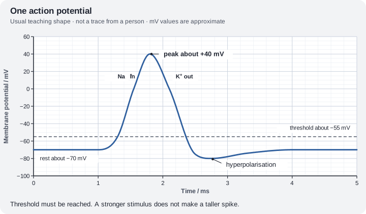
A stimulus opens some voltage-gated sodium channels. Sodium ions diffuse in, down their electrochemical gradient, and the inside becomes less negative. This is depolarisationA change in membrane potential that makes the inside less negative, as Na⁺ ions enter.. If the potential reaches a threshold, about −55 mV in the usual teaching account, many more sodium channels open. The inside rises rapidly, often to about +40 mV. That spike is the action potential. Sodium permeability then falls because those channels close.
Potassium restores the sign
Voltage-gated potassium channels open as the spike develops. Potassium ions diffuse out. The inside becomes negative again. This is repolarisationThe return of the membrane potential towards the resting value, as K⁺ ions leave.. The potassium channels are slow to close, so the potential briefly becomes more negative than rest. That is hyperpolarisationWhen the membrane potential becomes more negative than the resting potential.. The pump keeps the gradients from running down over many impulses. It is too slow to be the spike itself.
Once threshold is reached, the spike has a fixed size. A stronger stimulus does not make a taller action potential. That is all-or-nothing. Strength is coded by how often spikes are produced. During the spike and just after it, that patch of membrane cannot fire again. The refractory period is why the impulse does not reverse: the membrane behind the spike is unresponsive, while the membrane ahead can still be depolarised by local currents.
Myelin lets the current jump
In a myelinatedDescribes an axon wrapped in a myelin sheath. Impulses travel faster along it. axon, insulation forces the local current to the next node of RanvierA gap in the myelin sheath, where the axon membrane is exposed and action potentials can form.. Voltage-gated channels are concentrated at the nodes. The action potential is regenerated there and appears to jump from node to node. This is saltatory conductionConduction along a myelinated axon in which the action potential jumps from one node of Ranvier to the next, making it faster.. It is faster than continuous conduction along an unmyelinated axon of the same diameter. A wider axon is also faster, because there is less resistance to local current. Do not swap those two reasons.
Damaged myelin slows or blocks this jumping. The specification cell for peripheral myelin is the Schwann cellA cell that wraps around an axon to form the myelin sheath.. You do not need a disease name to explain 8.5.
| Stage | Permeability | Ion movement |
|---|---|---|
| Rest | Some K⁺ leak | Inside negative |
| Depolarisation | Na⁺ channels open | Na⁺ in |
| Repolarisation | K⁺ channels open, Na⁺ channels shut | K⁺ out |
| After the spike | Pump continues | Na⁺ out, K⁺ in, using ATP |
Make your reasoning visible
Sodium in, potassium out
Do not reverse them. The spike is sodium entry. Recovery of the negative inside is potassium exit.
Jump, do not flow through myelin
The impulse is regenerated at nodes. Myelin is not a pipe full of ions.
Avoid this misconception
The claim: The sodium-potassium pump causes the rapid rise of the spike, and a bigger stimulus makes a bigger spike.
The correction: The rapid rise is sodium diffusing in through opened channels. The pump maintains gradients. Above threshold, spike height stays the same and frequency can rise.
How a neurone sends a signal
A neurone sends a message as a brief electrical change along its axon. Channels in the membrane open and close, letting sodium and potassium ions move in and out. That change, the action potential, passes along the axon like a wave.
What the model shows
A tiny patch of axon membrane and its voltage over a few milliseconds when you stimulate it. A second view races an impulse along two kinds of axon.
What to look for
Which ions move, and in which direction, as the voltage rises and falls. What happens with a weak stimulus, a strong one, two close together, and one that keeps going. Which axon wins the race.
What it tells us
The rise is sodium flowing in and the fall is potassium flowing out. A spike happens fully or not at all. Just after a spike the membrane cannot fire again. A stronger stimulus gives more spikes, not bigger ones. Myelin makes the impulse jump between gaps, so it travels faster.
Be careful what you claim
The voltages are typical teaching values. Ion numbers are drawn small, and everything is slowed down a lot so you can see it.
Try it in the model: press Stimulate at strength 2, then at strength 5, and compare the two traces.
The action potential
Stimulate a patch of axon membrane and watch the Na⁺ and K⁺ channels and the voltage trace. Then race an impulse along an unmyelinated and a myelinated axon.
What you see
- A small patch of axon membrane with voltage-gated Na⁺ channels (orange) and K⁺ channels (purple). Open channels let ions through; an inactivated Na⁺ channel is plugged.
- Next to it, a live trace of the membrane potential (mV) with rest, threshold and peak lines, and the stimulus underneath.
- In the Conduction view, an unmyelinated and a myelinated axon of the same diameter, side by side.
How to use it
- Choose a stimulus: one pulse, two pulses with a gap, or a held stimulus. Set the strength, then press Stimulate.
- The last trace stays faint so you can compare. Pause, slow down or step through a spike.
- In the Conduction view, set the axon diameter and press Race. Each race is added to the results table.
What it shows
- Depolarisation is Na⁺ entry; repolarisation is K⁺ exit; K⁺ channels close slowly, so there is a brief hyperpolarisation.
- Threshold, all-or-nothing spikes, the refractory period, and frequency coding of stimulus strength.
- Myelinated axons conduct faster (saltatory conduction), and wider axons conduct faster.
Limits of the model
- The membrane uses the Hodgkin–Huxley equations for a squid axon, shifted to the usual teaching values (rest −70 mV, threshold about −55 mV, peak about +40 mV). Real neurones vary.
- Channel and ion numbers are tiny and drawn for clarity; very few ions actually cross, so concentrations hardly change in one spike. The Na⁺/K⁺ pump keeps the gradients over many impulses and is not drawn here.
- In a fast train each spike starts while the membrane is still partly refractory, so later spikes are a little smaller. Real neurones usually fire more slowly than this model.
- Race speeds use typical formulas (about 1.1√d m/s unmyelinated, 6d m/s myelinated) and are slowed down a lot. Nodes are not drawn to scale.
Guided tour · step 1 of 9
- In Stimulus, just below these steps, set Strength (0–10) to 2.
- Press Stimulate and watch the trace.
A few Na⁺ channels open and the inside becomes a little less negative, but it does not reach threshold (about −55 mV), so it falls back.
Stimulus
Questions
Final check: put it in order
What to do
- Reorder the cards below: drag a card (on a touch screen, drag its number), tap a card then tap where it goes, or use the ↑ and ↓ arrows at its right.
- Press Check under the cards to mark each one green (right place) or red; the count shows how many are right.
- Stuck? Hint first outlines the first wrong place, then gives a clue, then places that step for you.
- Shuffle mixes the cards again; Start again returns them to the starting order. Keyboard: Space picks up, arrow keys move, Space drops.
Put the events of one action potential in order, from rest back to rest.
1 of 6 in the right place
Values are typical; they vary between neurones. Saltatory conduction along a myelinated axon is in the race view of the model.
Check your understanding
Try each question before opening the suggested answer.
Check your understanding (continued)
Practice question
Retrieval and explanation · suggested answer
Which ion enters the axon during depolarisation, and why can it enter?
Show suggested answer
Sodium. Voltage-gated sodium channels open, and sodium diffuses in down its electrochemical gradient.
Practice question
Retrieval and explanation · suggested answer
How does the inside become negative again?
Show suggested answer
Sodium channels close and potassium channels open, so potassium ions diffuse out.
Practice question
Retrieval and explanation · suggested answer
Why does the impulse travel in only one direction along an axon?
Show suggested answer
The region just behind the spike is refractory and cannot be depolarised again immediately. Local currents therefore stimulate only the resting membrane ahead.
Practice question
Retrieval and explanation · suggested answer
What is saltatory conduction?
Show suggested answer
The action potential jumps from one node of Ranvier to the next in a myelinated axon, which is faster than continuous conduction.
Take the idea further
Apply the same specification knowledge to an unfamiliar problem. Any extra assumptions are supplied in the question.
Stretch question
A drug keeps voltage-gated sodium channels closed and does not affect the pump. Predict the effect on action potentials. Would the resting potential have to become zero immediately? Assume the gradients are still present at the moment the drug binds.
Show a hint
The spike needs the channels. Rest is mostly the gradients that already exist.
Explore the reasoning
New action potentials cannot start, because depolarisation needs sodium entry through those channels. The resting potential does not fall to zero at once, because the existing ion gradients and potassium permeability remain, and the pump can still run. Over a long time, unused gradients would change, but the immediate effect is loss of spikes. This is the lidocaine idea, without a dose.
Recall this lesson
Lesson recall questions
Question 1 of 5: State which ions enter the axon during depolarisation of an action potential.
Action potential along an axon
Practice question
Retrieval and explanation · suggested answer
State which ions enter the axon during depolarisation of an action potential.
Lesson: 8A 3: The Neurones in Action
Try this from memory first.
Put your biology to work
Explore where these ideas appear in the world. Enrichment, not extra required recall.
A receptor potential is not yet this spike
8A · The Neurones in ActionThe National Eye Institute says photoreceptors turn light into electrical signals. In a rod, that first electrical event is a hyperpolarisation, taught in the sensory lesson, not the…Specification 8.4Read & use the scienceClose article
Why this matters
The National Eye Institute says photoreceptors turn light into electrical signals. In a rod, that first electrical event is a hyperpolarisation, taught in the sensory lesson, not the sodium spike drawn here. Action potentials appear later, in the optic neurones. Use the eye page as a warning not to give every cell the same curve. 8.4 and 8.5: sodium and potassium permeabilities make the spike, and myelin makes it saltatory.
Back to the specification
Pearson Edexcel IAL Biology · Issue 2, February 2021 · Unit 5, pp. 35–38. The application extends beyond required recall.
- 8.4 Action potentials and sodium/potassium permeability.
Use the science
Why is “light opens sodium channels in the rod, like an axon” the wrong transfer of this lesson?
Explore a possible response
In an axon, sodium entry depolarises and makes the spike. In a rod, light leads to sodium channels closing and the cell hyperpolarises. The optic neurone is where the action-potential story applies.
Explore the source
- National Eye Institute: how the eyes workUS National Eye Institute
A local anaesthetic tests the sodium step
8A · The Neurones in ActionThe National Cancer Institute drug dictionary describes lidocaine as interacting with voltage-gated sodium channels and reducing the transient rise in membrane permeability to sodium. That is the rising…Specification 8.4Read & use the scienceClose article
Why this matters
The National Cancer Institute drug dictionary describes lidocaine as interacting with voltage-gated sodium channels and reducing the transient rise in membrane permeability to sodium. That is the rising phase of the action potential in 8.4. Blocking it can prevent an impulse from passing through the treated nerve, which is useful in local anaesthesia. The drug is an application, not a new version of the resting-potential diagram. Sodium-channel block does not mean potassium channels have vanished or that every sensory receptor is inactive. A nerve can detect a stimulus yet fail to conduct the resulting signal onward.
Back to the specification
Pearson Edexcel IAL Biology · Issue 2, February 2021 · Unit 5, pp. 35–38. The application extends beyond required recall.
- 8.4 Action potentials and sodium/potassium permeability.
Use the science
Why can a receptor still detect a stimulus while lidocaine prevents the person from feeling it?
Explore a possible response
Detection at the receptor and propagation along the sensory axon are distinct steps. Lidocaine blocks sodium-dependent action potentials, so the signal may not reach the CNS.
Explore the source
- Lidocaine hydrochloride: drug definitionUS National Cancer Institute