
What you need to know
- 8.2The nervous system connects a stimulus to a response by an effector.
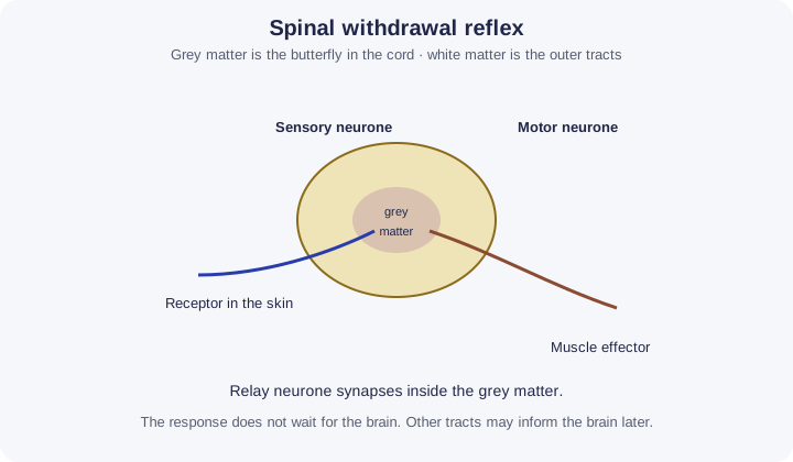
- 8.3A spinal reflex arcThe pathway of a reflex: receptor, sensory neurone, relay neurone, motor neurone, effector., including grey matter and white matter.
The pupil reflex is not spinal. It is with synapses in 8A 6. A knee-jerk reflex may have no relay neurone. The worked example here is the three-neurone withdrawal reflex the specification list describes.
By the end, you should be able to…
- Order receptor, sensory neuroneA neurone that carries impulses from a receptor to the central nervous system., relay, motor neuroneA neurone that carries impulses from the central nervous system to an effector, such as a muscle. and effector.
- Place the relay synapse in the grey matter.
- Explain why this response does not wait for a decision in the brain.
Build your understanding
From stimulus to response
A stimulus is a change in the environment. A receptor detects it and a sensory neuroneA neurone that carries impulses from a receptor to the central nervous system. carries impulses into the spinal cord. A relay neuroneA neurone in the central nervous system that carries impulses from sensory neurones to motor neurones. connects that sensory neurone to a motor neuroneA neurone that carries impulses from the central nervous system to an effector, such as a muscle.. The motor neurone carries impulses to an effector. The effector responds: a muscle contracts or a gland secretes. That chain is how the nervous system makes an effector answer a stimulus.
The response in a reflex is rapid and automatic. You do not choose it in the cerebral hemispheres first. That is useful when the stimulus is damaging, such as heat on the skin. The hand withdraws before you have finished thinking that it hurts.
Grey matter and white matter
In the spinal cord the grey matter is the central region. It contains cell bodies and the synapses of the reflex. The relay neurone lies here. White matter is outside the grey matter in the cord. It is pale because of myelin on the axons that run up and down the cord as tracts.
A sensory neurone enters through the dorsal root. Its cell body is in the dorsal root ganglion, in the white-matter side of the pathway but outside the cord itself. The motor neurone leaves through the ventral root. If a diagram is given, use dorsal and ventral rather than guessing left and right on the page.
The brain is informed, not required
Other neurones can carry impulses up the white-matter tracts to the brain, so you feel the pain. Those tracts are not the reflex arcThe pathway of a reflex: receptor, sensory neurone, relay neurone, motor neurone, effector.. Cutting the connection to the brain does not, by itself, abolish a spinal reflex. Damage to the sensory or motor neurone of the arc does.
Do not draw the pupil here. Iris muscles are controlled through the brain, not through a spinal relay. And if a question shows only two neurones, do not invent a relay that is not there.
Make your reasoning visible
Five words in order
Receptor, sensory neuroneA neurone that carries impulses from a receptor to the central nervous system., relay neuroneA neurone in the central nervous system that carries impulses from sensory neurones to motor neurones., motor neuroneA neurone that carries impulses from the central nervous system to an effector, such as a muscle., effector. Then say the synapse is in the grey matter.
White is tracts
White matter is myelinated axons running as pathways. Grey matter is the processing core of the cord.
Avoid this misconception
The claim: A reflex travels to the brain and back, and grey matter is the myelin.
The correction: A spinal reflex is completed in the cord. Grey matter contains the cell bodies and synapses. Myelin is what makes the surrounding white matter look white.
Put the reflex arc in order
A spinal reflex is fast because the route is fixed and short: it passes through the spinal cord, not the conscious brain. Order the route.
Order the steps
What to do
- Reorder the cards below: drag a card (on a touch screen, drag its number), tap a card then tap where it goes, or use the ↑ and ↓ arrows at its right.
- Press Check under the cards to mark each one green (right place) or red; the count shows how many are right.
- Stuck? Hint first outlines the first wrong place, then gives a clue, then places that step for you.
- Shuffle mixes the cards again; Start again returns them to the starting order. Keyboard: Space picks up, arrow keys move, Space drops.
Your hand touches a hot plate. Put the reflex arc in order.
0 of 6 in the right place
Some reflexes (for example the knee jerk) have no relay neurone. The impulse to the brain, which makes you feel the pain afterwards, is not part of the arc.
Check your understanding
Try each question before opening the suggested answer.
Check your understanding (continued)
Practice question
Retrieval and explanation · suggested answer
List, in order, the five parts of a three-neurone spinal reflex from stimulus to response.
Show suggested answer
Receptor, sensory neurone, relay neurone, motor neurone, effector.
Practice question
Retrieval and explanation · suggested answer
Where in the spinal cord is the relay synapse?
Show suggested answer
In the grey matter.
Practice question
Retrieval and explanation · suggested answer
Why is the withdrawal reflex faster than a voluntary movement that you think about first?
Show suggested answer
The arc uses few synapses in the cord and does not wait for processing in the cerebral hemispheres.
Practice question
Retrieval and explanation · suggested answer
What is white matter made of, in this context?
Show suggested answer
Myelinated axons running in tracts.
Take the idea further
Apply the same specification knowledge to an unfamiliar problem. Any extra assumptions are supplied in the question.
Stretch question
A person’s spinal reflex still withdraws the hand from heat, but they report no sensation. Assume the reflex neurones are intact and a tract to the brain is not. Explain both observations. Why is this not evidence that the brain starts the reflex?
Show a hint
Separate the arc from the ascending tract.
Explore the reasoning
The local arc still runs from receptor to muscle, so the hand withdraws. Sensation needs impulses to reach the brain, and that tract is the part assumed to be damaged. The reflex happening without sensation shows the brain is not required to start it.
Recall this lesson
Lesson recall questions
Question 1 of 5: Name the component that detects a stimulus and converts it into a nerve impulse.
From stimulus to effector
Practice question
Retrieval and explanation · suggested answer
Name the component that detects a stimulus and converts it into a nerve impulse.
Lesson: 8A 2: How the Nervous System Works
Try this from memory first.
Put your biology to work
Explore where these ideas appear in the world. Enrichment, not extra required recall.
A receptor starts the pathway the cord then finishes
8A · How the Nervous System WorksThe National Eye Institute describes photoreceptors turning light into electrical signals. A spinal reflex uses the same idea of a receptor, then neurones, then an effector, but the…Specification 8.2Read & use the scienceClose article
Why this matters
The National Eye Institute describes photoreceptors turning light into electrical signals. A spinal reflex uses the same idea of a receptor, then neurones, then an effector, but the worked example is the cord, not the eye. Do not move the pupil reflex into the grey matter of the spine. 8.2 and 8.3: a stimulus leads to an effector response, and a spinal reflex does that through grey matter. A behaviour or symptom is the output of a pathway, not a direct recording of ion movement in one axon or synapse.
Back to the specification
Pearson Edexcel IAL Biology · Issue 2, February 2021 · Unit 5, pp. 35–38. The application extends beyond required recall.
- 8.2 Nervous responses from stimulus to effector.
Use the science
Why is blinking in bright light a poor diagram for 8.3?
Explore a possible response
8.3 is a spinal arc, with grey and white matter of the cord. The pupil and many eye reflexes are coordinated in the brain. Use a withdrawal from the skin if you need the spinal list.
Explore the source
- National Eye Institute: how the eyes workUS National Eye Institute
After spinal injury, signal direction matters
8A · How the Nervous System WorksNINDS describes the spinal cord as the route that sends and receives signals between brain and body. An injury can interrupt sensory information travelling in and motor commands…Specification 8.2Read & use the scienceClose article
Why this matters
NINDS describes the spinal cord as the route that sends and receives signals between brain and body. An injury can interrupt sensory information travelling in and motor commands travelling out, though the effects depend strongly on its location and extent. This applies the 8.2 stimulus-to-effector chain to rehabilitation: a therapist needs to know whether the difficulty is sensing a stimulus, processing it centrally or activating a muscle. A visible movement does not by itself show that every part of the original pathway is intact; local reflex circuits can still act in some circumstances.
Back to the specification
Pearson Edexcel IAL Biology · Issue 2, February 2021 · Unit 5, pp. 35–38. The application extends beyond required recall.
- 8.2 Nervous responses from stimulus to effector.
Use the science
Why would the loss of voluntary movement below a spinal injury not prove that every local reflex arc below it is destroyed?
Explore a possible response
Voluntary commands from the brain may be interrupted while sensory, relay and motor neurones in a local spinal circuit remain connected. Reflex behaviour needs separate testing.
Explore the source
- Spinal cord injuryUS National Institute of Neurological Disorders and Stroke