
What you need to know
- 8.1Structure and function of sensory, relay and motor neuroneA neurone that carries impulses from the central nervous system to an effector, such as a muscle., including Schwann cellA cell that wraps around an axon to form the myelin sheath. and myelination.
Saltatory conduction is 8A 3. The synapse is 8A 6. Schwann cells are the peripheral myelin cells named in the specification. Do not call every glial cell a Schwann cell.
By the end, you should be able to…
- Describe the direction of an impulse in each of the three neurones.
- Place the cell body of a sensory neuroneA neurone that carries impulses from a receptor to the central nervous system. and of a motor neurone.
- State what a Schwann cell does and what a node of RanvierA gap in the myelin sheath, where the axon membrane is exposed and action potentials can form. is.
Build your understanding
Three cells, one direction each
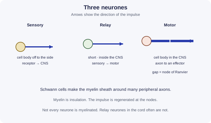
A sensory neuroneA neurone that carries impulses from a receptor to the central nervous system. carries impulses from a receptor toward the central nervous systemThe brain and spinal cord.. Its cell body sits to the side of the fibre, often in a ganglion just outside the spinal cord. A long fibre brings the message in from the receptor, and a shorter fibre enters the cord.
Three cells, one direction each (continued)
A relay neurone is entirely inside the central nervous system. It passes the impulse from a sensory neurone to a motor neuroneA neurone that carries impulses from the central nervous system to an effector, such as a muscle., or on to other relay neuroneA neurone in the central nervous system that carries impulses from sensory neurones to motor neurones.. A motor neurone carries impulses out to an effector, a muscle or a gland. Its cell body is inside the central nervous system and its long axon runs to the effector. Dendrites receive. The axon carries the impulse away from the cell body.
Myelin is insulation, not the impulse
In the peripheral nervous systemAll the nerves outside the brain and spinal cord., Schwann cellA cell that wraps around an axon to form the myelin sheath. wrap many times around an axon. The wrapping is the myelin sheathA fatty insulating layer around some axons, formed by Schwann cells., mostly lipid, so it insulates the membrane. Gaps between neighbouring Schwann cells are node of RanvierA gap in the myelin sheath, where the axon membrane is exposed and action potentials can form.. The membrane is bare at a node.
Myelination matters because the impulse can jump from node to node. That is saltatory conductionConduction along a myelinated axon in which the action potential jumps from one node of Ranvier to the next, making it faster., taught with the action potentialA rapid reversal of the potential difference across a neurone membrane, caused by Na⁺ ions flowing in, then K⁺ ions flowing out.. Here you need the structure: Schwann cell, sheath, node. Many relay neurones in the central nervous system are not wrapped by Schwann cells. Do not draw myelin on every neurone by habit.
Function follows the wiring
Receptors and effectors are not neurones. A receptor detects the stimulus and starts an impulse in the sensory neurone. The effector does the response when the motor neurone arrives. Mixing those words loses the mark.
The cell bodies contain the nucleusThe organelle that contains the cell’s chromosomes, surrounded by a nuclear envelope with pores. and most of the organelleA structure inside a cell with a particular function, such as a mitochondrion or ribosome.. An axon can be long because the impulse is a membrane event, not a flow of cytoplasmEverything inside the cell surface membrane except the nucleus: the fluid cytosol and the organelles. down the tube.
| Neurone | Cell body | Carries the impulse |
|---|---|---|
| Sensory | To the side, often in a ganglion | Receptor toward the CNS |
| Relay | Inside the CNS | Sensory neurone toward a motor neurone |
| Motor | Inside the CNS | CNS toward an effector |
Make your reasoning visible
Toward or away
Sensory in, motor out. Relay stays in the CNS.
Schwann, then node
Name the cell that makes the sheath, then the gap where the membrane is exposed.
Avoid this misconception
The claim: All three neurones look the same, and myelin is a blood vessel along the axon.
The correction: The cell-body position differs. Myelin is a lipid wrap made by Schwann cellA cell that wraps around an axon to form the myelin sheath. in the peripheral nervous systemAll the nerves outside the brain and spinal cord.. It insulates. It is not a vessel.
Check your understanding
Try each question before opening the suggested answer.
Practice question
Retrieval and explanation · suggested answer
Where is the cell body of a sensory neurone, and in which direction does its impulse travel?
Show suggested answer
The cell body is to the side of the fibre, often in a ganglion outside the cord. The impulse travels from a receptor toward the CNS.
Practice question
Retrieval and explanation · suggested answer
What is the function of a motor neurone?
Show suggested answer
It carries impulses from the CNS to an effector such as a muscle or a gland.
Practice question
Retrieval and explanation · suggested answer
What is a node of Ranvier?
Show suggested answer
A gap in the myelin sheath between Schwann cells, where the axon membrane is exposed.
Take the idea further
Apply the same specification knowledge to an unfamiliar problem. Any extra assumptions are supplied in the question.
Stretch question
A drawing shows a long axon from the spinal cord to a muscle, with a cell body at the cord end and no side branch for a cell body. A student labels it sensory because it is long. What should the label be, and which 8.1 feature is still missing if you also want to discuss speed?
Show a hint
Direction and cell-body position first. Speed needs the sheath.
Explore the reasoning
It is a motor neurone: cell body in the CNS, axon to a muscle. Length does not decide the type. The drawing has not shown Schwann cells or nodes, so you cannot yet discuss saltatory conduction. Those structures are the myelination part of 8.1.
Recall this lesson
Lesson recall questions
Question 1 of 5: Describe, in order, how a Schwann cell myelinated an axon.
Neurone types and myelin
Practice question
Retrieval and explanation · suggested answer
Describe, in order, how a Schwann cell myelinated an axon.
Lesson: 8A 1: The Structure of Neurones
Build the sequence from memory first. Then reveal the model order.
Tap the steps in the order you recall. Tap a placed step to send it back.
- 01Next step
- 02Waiting
- 03Waiting
Tap to place
Put your biology to work
Explore where these ideas appear in the world. Enrichment, not extra required recall.
Light becomes a signal only after a receptor cell
8A · The Structure of NeuronesThe National Eye Institute describes the retina as a light-sensitive layer where photoreceptors turn light into electrical signals. That is the start of a sensory pathway. The page…Specification 8.1Read & use the scienceClose article
Why this matters
The National Eye Institute describes the retina as a light-sensitive layer where photoreceptors turn light into electrical signals. That is the start of a sensory pathway. The page does not name Schwann cells or the three neurone types. The specification structure is still yours to learn. Rod detail is a later lesson. 8.1: sensory neurones carry impulses that began at a receptor. Motor neurones end at an effector. A behaviour or symptom is the output of a pathway, not a direct recording of ion movement in one axon or synapse.
Back to the specification
Pearson Edexcel IAL Biology · Issue 2, February 2021 · Unit 5, pp. 35–38. The application extends beyond required recall.
- 8.1 Sensory, relay and motor neurones, Schwann cells and myelin.
Use the science
Why is “the eye is a motor neurone” a category error?
Explore a possible response
The eye contains receptors. A sensory neurone carries the impulse inward. A motor neurone would carry an impulse out to an effector. The NEI sentence is about detection, not about myelin.
Explore the source
- National Eye Institute: how the eyes workUS National Eye Institute
Myelin damage changes a network, not just one cable
8A · The Structure of NeuronesThe US neurological institute describes multiple sclerosis as immune injury to myelin in the central nervous system. Myelin insulates parts of an axon and supports rapid signal conduction,…Specification 8.1Read & use the scienceClose article
Why this matters
The US neurological institute describes multiple sclerosis as immune injury to myelin in the central nervous system. Myelin insulates parts of an axon and supports rapid signal conduction, so damage can disrupt communication across sensory and motor pathways. Your 8.1 diagram includes Schwann cells, which myelinate peripheral axons; central myelin is made by oligodendrocytes. That distinction matters because MS affects the central system. Symptoms depend on which pathways are injured, not on one fixed sketch of a damaged sheath. Clinicians combine history, examination and imaging because the biology occurs in a network, not in an isolated textbook neurone.
Back to the specification
Pearson Edexcel IAL Biology · Issue 2, February 2021 · Unit 5, pp. 35–38. The application extends beyond required recall.
- 8.1 Sensory, relay and motor neurones, Schwann cells and myelin.
Use the science
Why might myelin damage produce a sensory problem in one person and a movement problem in another?
Explore a possible response
The effect depends on where the damage lies. Disrupting sensory pathways changes information reaching the CNS; disrupting motor pathways changes signals sent to effectors.
Explore the source
- Multiple sclerosis: hope through researchUS National Institute of Neurological Disorders and Stroke