
What you need to know
- 7.11sliding filamentThe model of muscle contraction: actin filaments slide between myosin filaments, so each sarcomere shortens. theory: actinThe protein of the thin filaments in muscle. Myosin heads bind to it., myosinThe protein of the thick filaments in muscle. Its heads bind to actin and pull it, using ATP., troponinA protein on actin filaments. When Ca²⁺ binds to troponin, it moves tropomyosin off the myosin-binding sites., tropomyosinA protein strand that covers the myosin-binding sites on actin when the muscle is relaxed., calcium ions, ATPAdenosine triphosphate: the molecule that releases energy for cell processes when it is hydrolysed to ADP and Pi. and ATPaseAn enzyme that hydrolyses ATP to ADP and Pi, releasing energy. The myosin head is an ATPase..
The A band, I band and H zone explain the evidence that filaments slide. They are part of describing the sarcomere, not extra proteins. No named neuromuscular transmitter is required here; synapses are Topic 8.
By the end, you should be able to…
- Describe how calcium ions expose myosin-binding sites on actin.
- Outline the cross-bridge cycle and the two uses of ATP.
- State which parts of the sarcomereThe unit of a myofibril between two Z lines. It shortens when the muscle contracts. shorten and which do not.
Build your understanding
Calcium uncovers the binding site
An impulse reaches the fibre, T-tubules carry it inward, and the sarcoplasmic reticulum releases calcium ions into the sarcoplasmThe cytoplasm of a muscle fibre.. Calcium ions bind to troponinA protein on actin filaments. When Ca²⁺ binds to troponin, it moves tropomyosin off the myosin-binding sites.. Troponin changes shape and pulls tropomyosinA protein strand that covers the myosin-binding sites on actin when the muscle is relaxed. away from the myosin-binding sites on actinThe protein of the thin filaments in muscle. Myosin heads bind to it.. Until that happens, tropomyosin blocks the sites and cross-bridges cannot form.
When calcium ions are pumped back into the sarcoplasmic reticulum, tropomyosin covers the sites again and the fibre can relax. That pumping uses ATPAdenosine triphosphate: the molecule that releases energy for cell processes when it is hydrolysed to ADP and Pi.. Relaxation is not passive in the sense of “no energy anywhere”.
The cross-bridge cycle
The head of myosinThe protein of the thick filaments in muscle. Its heads bind to actin and pull it, using ATP. is an ATPaseAn enzyme that hydrolyses ATP to ADP and Pi, releasing energy. The myosin head is an ATPase.. hydrolysisA reaction that breaks a covalent bond using a molecule of water. of ATP to ADPAdenosine diphosphate. It is formed when ATP is hydrolysed, and is phosphorylated to make ATP again. and phosphate cocks the head into a high-energy position. The cocked head binds an exposed site on actin, forming a cross-bridge. The head then tilts in the power stroke, sliding actin toward the centre of the sarcomereThe unit of a myofibril between two Z lines. It shortens when the muscle contracts., and ADP and phosphate are released.
A new ATP molecule binds the myosin head, which makes it detach from actin. Without that ATP the bridge stays locked. Hydrolysis of the new ATP cocks the head again. As long as calcium ions and ATP are present, the cycle repeats. Many heads work asynchronously so the filament does not slip back as a sheet.
What shortens is the sarcomere, not the myosin
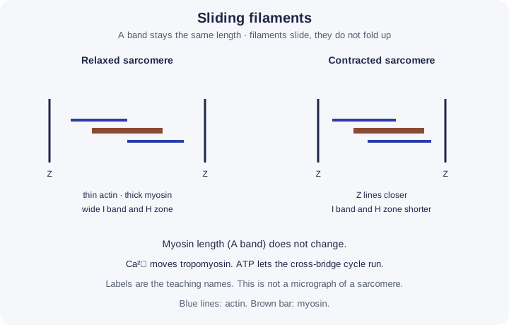
Filaments slide. They do not fold up as the main event. The A band, which is the length of the myosin filament, stays the same. The I band, actin that does not overlap myosin, shortens. The H zone, myosin that does not overlap actin, shortens. Z lines move closer, so the sarcomere shortens and the fibre can shorten.
If every sarcomere in a fibre shortens together, the fibre pulls on its tendonTough, inelastic connective tissue that attaches a muscle to a bone.. That is how 7.11 meets 7.9. The theory does not say the muscle pushes.
Follow the process
Impulse along the T-tubules → the sarcoplasmic reticulum releases calcium ions → calcium binds troponin and tropomyosin moves off the myosin-binding sites on actin → the myosin head, already cocked by hydrolysis of ATP, binds and forms a cross-bridge → the head tilts in the power stroke and actin slides toward the centre of the sarcomere → a new ATP binds and the head detaches → that ATP is hydrolysed and the head is cocked again. Calcium pumped back into the reticulum lets tropomyosin cover the sites, so the fibre can relax. The A band stays the same length. The I band and the H zone shorten. The filaments slide. They do not fold up, and calcium is not the energy source.
| Region | What it spans | During contraction |
|---|---|---|
| A band | Length of myosin | Stays the same |
| I band | Actin only | Shortens |
| H zone | Myosin only | Shortens |
| Z to Z | One sarcomere | Shortens |
Make your reasoning visible
Build the explanation
X happens because Y, leading to Z. X: the sarcomereThe unit of a myofibril between two Z lines. It shortens when the muscle contracts. shortens. Y: myosinThe protein of the thick filaments in muscle. Its heads bind to actin and pull it, using ATP. heads pull actinThe protein of the thin filaments in muscle. Myosin heads bind to it. inward while tropomyosinA protein strand that covers the myosin-binding sites on actin when the muscle is relaxed. is held off the binding sites. Z: the Z lines move closer, but the myosin filament itself stays the same length, so the A band does not change. Incomplete: “The muscle contracts because calcium shortens myosin.” Improved: “Calcium exposes the binding sites. ATPAdenosine triphosphate: the molecule that releases energy for cell processes when it is hydrolysed to ADP and Pi. lets the myosin head detach and re-cock. The power stroke slides actin, so the sarcomere shortens without the filaments folding.”
Calcium, then ATP jobs
Calcium moves tropomyosin. ATP detaches the head and, after hydrolysisA reaction that breaks a covalent bond using a molecule of water., cocks it. ATPaseAn enzyme that hydrolyses ATP to ADP and Pi, releasing energy. The myosin head is an ATPase. is the myosin head.
A band is the constant one
If a mark-style question asks which band does not change, it is the A band.
Avoid this misconception
The claim: Myosin shortens and pushes actin, and calcium is the energy source.
The correction: Myosin heads pull actin so the filaments slide. ATP supplies the energy. Calcium ions only expose the binding sites.
How a muscle shortens without its filaments shrinking
Under an electron microscope, a contracted muscle fibre shows bands, and some of them get shorter while one stays exactly the same. The sliding filament theory explains why. The model below lets you watch it happen in one sarcomere.
What the model shows
One sarcomere, the tiny repeating unit inside a muscle fibre. Thin actin filaments are fixed to the Z lines at each end. Thick myosin filaments sit in the middle, covered in small heads that can grab actin.
What to look for
Watch the myosin heads grab actin, tilt and let go, and see which bands get shorter. Keep an eye on the A band reading: it never changes. Slow motion helps you follow a single head.
What it tells us
Muscles shorten because actin and myosin slide past each other, not because the filaments shrink. Calcium ions are the on switch: they uncover the binding sites on actin. ATP supplies the energy that lets each head let go and re-cock.
Be careful what you claim
It is a simplified drawing with far fewer heads than a real filament, and the speeds are chosen so you can see them. It does not show how much force the muscle makes or what it is pulling against.
Try it in the model: release calcium and watch the I band and H zone shrink. Then switch the ATP supply off and see why the muscle locks.
Slide the filaments
One sarcomere, schematic. Release calcium ions to uncover the binding sites, watch myosin heads cycle on actin, and read which bands change. Then take the ATP away. Rates are illustrative.
What you see
- One sarcomere between two Z lines: blue actin (thin) filaments with tropomyosin, purple myosin (thick) filaments with heads.
- Head colour shows its state: orange cocked (ADP + Pi), red after the power stroke, green with a new ATP bound. Yellow dots are free Ca²⁺.
- Brackets for the A band, the I band halves, the H zone and the sarcomere, with a trace of sarcomere length over time.
How to use it
- Stimulate to release Ca²⁺; stop to let it be pumped back.
- Switch the ATP supply off to see rigor. Use slow motion to follow single heads.
- “Relaxed fibre, no ATP” sets up the lesson’s stretch question.
What it shows
- Calcium ions expose binding sites by moving tropomyosin; they are not the energy source.
- The cross-bridge cycle and the two roles of ATP at the head: detaching and re-cocking.
- Filaments slide: the A band stays the same while the I band, H zone and sarcomere shorten.
Limits of the model
- Schematic: a few dozen heads stand for hundreds per filament; rates and step size are chosen for viewing, not measured.
- Shortening is unloaded, and relaxation uses a simple elastic return. Force, load and titin are not modelled.
- Lengths (A band 1.6 µm, actin 1.0 µm, rest 2.6 µm) are typical textbook values.
Guided tour · step 1 of 7
- In Controls, just below these steps, press Stimulate: release Ca²⁺.
- Watch the yellow Ca²⁺ ions appear in the simulator and the tropomyosin strand slide off the binding sites on actin.
An impulse makes the sarcoplasmic reticulum release Ca²⁺. Ca²⁺ binds troponin, which pulls tropomyosin away so the myosin-binding sites on actin are exposed.
Controls
Readings
Power strokes so far: 0. ATP molecules hydrolysed: 0. Start: A 1.60, I 1.00, H 0.60, sarcomere 2.60 µm.
Bands
Stretch
ATP’s jobs
Final check: put it in order
What to do
- Reorder the cards below: drag a card (on a touch screen, drag its number), tap a card then tap where it goes, or use the ↑ and ↓ arrows at its right.
- Press Check under the cards to mark each one green (right place) or red; the count shows how many are right.
- Stuck? Hint first outlines the first wrong place, then gives a clue, then places that step for you.
- Shuffle mixes the cards again; Start again returns them to the starting order. Keyboard: Space picks up, arrow keys move, Space drops.
An impulse reaches a muscle fibre. Put one cycle of contraction in order.
2 of 7 in the right place
When stimulation stops, Ca²⁺ is pumped back into the sarcoplasmic reticulum using ATP and tropomyosin covers the sites again. Many heads cycle out of step.
Check your understanding
Try each question before opening the suggested answer.
Check your understanding (continued)
Practice question
Retrieval and explanation · suggested answer
What does calcium do in the sliding filament mechanism?
Show suggested answer
It binds troponin, which moves tropomyosin and exposes myosin-binding sites on actin.
Practice question
Retrieval and explanation · suggested answer
State two roles of ATP in contraction or relaxation.
Show suggested answer
Hydrolysis cocks the myosin head. Binding of a new ATP detaches the head. Pumping calcium back into the sarcoplasmic reticulum also uses ATP. Any two.
Practice question
Retrieval and explanation · suggested answer
Which band stays the same length, and why?
Show suggested answer
The A band, because it is set by the length of the myosin filaments, which do not shorten.
Practice question
Retrieval and explanation · suggested answer
Why does a lack of ATP cause muscles to stay stiff?
Show suggested answer
Myosin cannot detach from actin without a new ATP, so cross-bridges remain locked.
Take the idea further
Apply the same specification knowledge to an unfamiliar problem. Any extra assumptions are supplied in the question.
Stretch question
A fibre is stimulated in a medium with calcium ions but no ATP. Predict whether cross-bridges can keep cycling, and whether tropomyosin can uncover the sites. Assume the reticulum can still release the calcium that is already stored.
Show a hint
Separate the switch from the fuel.
Explore the reasoning
Calcium can still bind troponin and move tropomyosin, so sites can be exposed. Without ATP the heads cannot complete the detach-and-cock cycle, so repeated sliding stops. Any bridges that form may lock. The assumption is that stored calcium can be released once; it does not invent a new energy source.
Recall this lesson
Lesson recall questions
Question 1 of 5: Describe, in order, how calcium ions allow myosin to bind actin.
Sliding filament theory
Practice question
Retrieval and explanation · suggested answer
Describe, in order, how calcium ions allow myosin to bind actin.
Lesson: 7B 4: How Muscles Contract
Build the sequence from memory first. Then reveal the model order.
Tap the steps in the order you recall. Tap a placed step to send it back.
- 01Next step
- 02Waiting
- 03Waiting
Tap to place
Put your biology to work
Explore where these ideas appear in the world. Enrichment, not extra required recall.
Soreness is not a cross-bridge diagram
7B · How Muscles ContractNHS information on muscle strains describes torn or overstretched muscle. It does not set out troponin, tropomyosin or ATP hydrolysis. Do not explain a strain by “the A…Specification 7.11Read & use the scienceClose article
Why this matters
NHS information on muscle strains describes torn or overstretched muscle. It does not set out troponin, tropomyosin or ATP hydrolysis. Do not explain a strain by “the A band got shorter”. The A band does not get shorter in normal contraction either. 7.11: sliding filaments, calcium uncovering binding sites, ATP and ATPase. An injury, symptom or whole-body performance result is not a direct image of a sarcomere or cardiac conduction pathway.
Back to the specification
Pearson Edexcel IAL Biology · Issue 2, February 2021 · Unit 5, pp. 35–38. The application extends beyond required recall.
- 7.11 Sliding filament contraction and the roles of calcium, ATP and ATPase.
Use the science
A friend says the A band shortens because the muscle is sore. What does the sliding filament evidence actually say about the A band?
Explore a possible response
The A band stays the same length because myosin filaments do not shorten. Soreness is not evidence about band length.
Explore the source
Why a stopped ATP supply stiffens muscle
7B · How Muscles ContractA molecular review of actin–myosin interaction explains a counterintuitive part of contraction: ATP binding helps a myosin head detach from actin, while ATP hydrolysis prepares another force-producing cycle.…Specification 7.11Read & use the scienceClose article
Why this matters
A molecular review of actin–myosin interaction explains a counterintuitive part of contraction: ATP binding helps a myosin head detach from actin, while ATP hydrolysis prepares another force-producing cycle. When ATP production ceases after death, many attachments can remain, contributing to rigor mortis. This is a striking application of 7.11, but a living muscle does not work by staying in permanent rigor. Calcium still controls access to actin, and repeated attachment and detachment generate movement. The example also links Topic 7 to forensic science without making post-mortem timing an extra requirement for the muscle lesson.
Back to the specification
Pearson Edexcel IAL Biology · Issue 2, February 2021 · Unit 5, pp. 35–38. The application extends beyond required recall.
- 7.11 Sliding filament contraction and the roles of calcium, ATP and ATPase.
Use the science
Why would removing ATP stop both repeated shortening and normal relaxation?
Explore a possible response
Myosin needs ATP binding to detach from actin and ATP hydrolysis to reset for another cycle. Without ATP, cross-bridges can remain attached and cycles cannot continue.
Explore the source
- The actin–myosin interaction in musclePeer-reviewed review via NIH PubMed Central