
What you need to know
- 7.12(i)Cardiac muscle is myogenicDescribes heart muscle that contracts by itself, without needing nerve impulses..
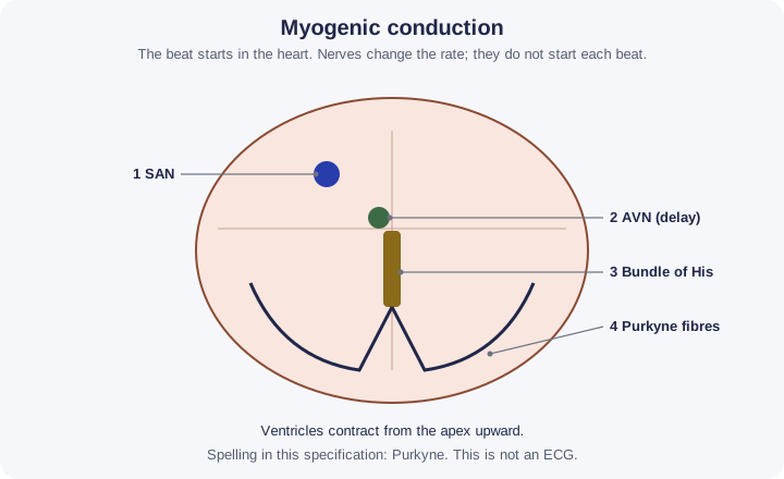
- 7.12(ii)The heartbeat is coordinated by the sinoatrial nodeThe pacemaker: a patch of tissue in the right atrium wall that sends out a wave of excitation across the atria., atrioventricular nodeTissue between the atria that delays the impulse briefly, so the atria finish emptying, then passes it to the bundle of His., bundle of HisConducting fibres in the septum that carry the impulse from the AVN down to the apex of the heart. and Purkyne fibresConducting fibres in the ventricle walls that spread the impulse upwards from the apex, so the ventricles contract from the bottom up..
- 7.12(iii)Electrocardiograms can help diagnose abnormal heart rhythms.
Use the spelling Purkyne. Nerves and adrenaline change the rate; they are the next control lesson, not the origin of each beat. An ECG in this course is not a diagnosis you make of a real person.
By the end, you should be able to…
- Define myogenic and name the nodes in order.
- Explain the delay at the atrioventricular node.
- Match a normal P wave, QRS complex and T wave to depolarisationA change in membrane potential that makes the inside less negative, as Na⁺ ions enter., without diagnosing a patient.
Build your understanding
The beat starts in the muscle
Cardiac muscle is myogenicDescribes heart muscle that contracts by itself, without needing nerve impulses.. The impulse that starts each heartbeat is generated in the muscle itself, not by a nerve from the brain. The sinoatrial nodeThe pacemaker: a patch of tissue in the right atrium wall that sends out a wave of excitation across the atria., the SAN, in the wall of the right atriumAn upper chamber of the heart. It receives blood from the veins and pumps it into the ventricle below., sets the pace. The impulse spreads through the atrial muscle, so the atria contract together from the top.
If the nerves to the heart are cut, the heart can still beat. The rate would no longer be adjusted to exercise in the normal way. Myogenic means the origin of the beat, not “nothing can influence it”.
Delay, then the apex
The impulse reaches the atrioventricular nodeTissue between the atria that delays the impulse briefly, so the atria finish emptying, then passes it to the bundle of His., the AVN. There is a short delay. That delay lets the atria finish emptying into the ventricleA lower chamber of the heart. Its thick muscular wall pumps blood into an artery. before the ventricles contract. The impulse then travels down the bundle of HisConducting fibres in the septum that carry the impulse from the AVN down to the apex of the heart. in the septumThe wall of tissue that separates the left and right sides of the heart. and out through the Purkyne fibresConducting fibres in the ventricle walls that spread the impulse upwards from the apex, so the ventricles contract from the bottom up. in the ventricle walls.
Because the Purkyne fibres carry the impulse to the apex and then through the walls, ventricular contraction squeezes blood upward into the arteryA vessel that carries blood away from the heart at high pressure. It has a thick wall with elastic fibres and smooth muscle.. A wave that started at the top of the ventricles and travelled down would be less effective. Spell the fibres Purkyne in this specification.
What an ECG shows
An electrocardiogram records electrical activity at the skin, not the sound of valveA flap of tissue that closes when pressure behind it is higher, so blood can flow in only one direction. and not the volume of blood. In a normal trace the P wave is atrial depolarisationA change in membrane potential that makes the inside less negative, as Na⁺ ions enter., the QRS complex is ventricular depolarisation, and the T wave is ventricular repolarisationThe return of the membrane potential towards the resting value, as K⁺ ions leave.. The gap after P matches the AVN delay.
A trace can show a rhythm that is very fast, very slow, or irregular, or electrical activity that is uncoordinated. That information can help a clinician diagnose an abnormal rhythm. It does not, in this course, tell you the cause, the treatment, or what a particular patient’s trace “must” mean. Do not diagnose yourself or anyone else from a textbook sketch.
| Event | Where | What you should say |
|---|---|---|
| SAN fires | Right atrium | Myogenic origin |
| AVN delay | Between atria and ventricles | Atria empty first |
| Bundle of His and Purkyne fibres | Septum and ventricle walls | Ventricles contract from the apex |
| P, QRS, T | ECG recording | Atrial depolarisation, ventricular depolarisation, ventricular repolarisation |
Make your reasoning visible
Order is the question
sinoatrial nodeThe pacemaker: a patch of tissue in the right atrium wall that sends out a wave of excitation across the atria., atrial muscle, atrioventricular nodeTissue between the atria that delays the impulse briefly, so the atria finish emptying, then passes it to the bundle of His. delay, bundle of HisConducting fibres in the septum that carry the impulse from the AVN down to the apex of the heart., Purkyne fibresConducting fibres in the ventricle walls that spread the impulse upwards from the apex, so the ventricles contract from the bottom up., ventricular muscle.
ECG is electrical
Do not describe the QRS complex as “the ventricleA lower chamber of the heart. Its thick muscular wall pumps blood into an artery. emptying”.
Avoid this misconception
The claim: The brain starts every heartbeat, and the AVN is the pacemaker.
The correction: The SAN is the pacemaker and the muscle is myogenicDescribes heart muscle that contracts by itself, without needing nerve impulses.. The AVN delays the impulse. Nerves only modify the rate.
Who tells your heart to beat?
Not your brain. Heart muscle is myogenic: each beat starts in a small patch of the heart itself, the sinoatrial node (SAN). From there an electrical wave spreads in a set order, so the atria contract first and the ventricles contract a moment later, squeezing blood up and out.
What the model shows
The heart with its conduction pathway, a wave of electrical activity spreading through the muscle, and a schematic ECG that records that activity at the skin.
What to look for
The order of the steps, the pause at the atrioventricular node (AVN), and which part of the ECG matches which step.
What it tells us
The SAN sets the pace, the AVN delay lets the atria empty first, and the Purkyne fibres make the ventricles contract from the bottom up. P, QRS and T on an ECG match atrial depolarisation, ventricular depolarisation and ventricular repolarisation.
Be careful what you claim
The ECG is a drawn sketch, not a real recording. The "No P waves" trace is fictional. Never use a textbook ECG to diagnose yourself or anyone else.
Try it in the model: press Play, then use Jump to: AVN delay and look at which parts of the heart are shaded yellow.
Control of the heartbeat and the ECG
Follow the electrical wave from the sinoatrial node to the Purkyne fibres, and match each step to the P wave, QRS complex and T wave.
What you see
- Left: the heart with its conduction pathway. The SAN is in the right atrium wall, the AVN between the atria and ventricles, the bundle of His in the septum and the Purkyne fibres in the ventricle walls.
- Yellow shading shows muscle that is depolarised. It spreads over the atria from the SAN, then over the ventricles from the apex upward.
- Right: a schematic ECG of the last 3 seconds, and the steps of one beat with the current step highlighted.
How to use it
- Play in slow motion or jump to a step. Change the AVN delay. Switch to the fictional "No P waves" trace.
What it shows
- The heartbeat is myogenic: it starts at the SAN without a nerve impulse.
- The AVN delay lets the atria empty before the ventricles contract, and the ventricles contract from the apex upward.
- P = atrial depolarisation, QRS = ventricular depolarisation, T = ventricular repolarisation.
Limits of the model
- The ECG is a drawn, schematic trace, not a recording from a person. Its sizes and timings are typical, not exact.
- The "No P waves" trace is fictional and for learning only. It is not a diagnosis, and you should not use it to judge anyone's heart.
- Nerves and adrenaline, which change the rate, are left out here; they are in 7B 7.
Guided tour · step 1 of 8
- In Playback, just below these steps, press Play and watch one beat.
- Follow the yellow wave on the heart and the highlighted row in the list.
The beat starts in the heart muscle itself, at the sinoatrial node (SAN). Cardiac muscle is myogenic.
Playback
Questions
Final check: put it in order
What to do
- Reorder the cards below: drag a card (on a touch screen, drag its number), tap a card then tap where it goes, or use the ↑ and ↓ arrows at its right.
- Press Check under the cards to mark each one green (right place) or red; the count shows how many are right.
- Stuck? Hint first outlines the first wrong place, then gives a clue, then places that step for you.
- Shuffle mixes the cards again; Start again returns them to the starting order. Keyboard: Space picks up, arrow keys move, Space drops.
Put one heartbeat in order, from the pacemaker to the ventricles contracting.
0 of 6 in the right place
A layer of non-conducting tissue between the atria and ventricles is why the impulse must pass through the AVN. Nerves from the brain change the rate but do not start each beat.
Check your understanding
Try each question before opening the suggested answer.
Check your understanding (continued)
Practice question
Retrieval and explanation · suggested answer
What does myogenic mean for cardiac muscle?
Show suggested answer
The heartbeat is started by the heart muscle itself, at the sinoatrial node, not by a nerve impulse from the brain.
Practice question
Retrieval and explanation · suggested answer
Why is there a delay at the AVN?
Show suggested answer
So the atria can finish contracting and empty blood into the ventricles before the ventricles contract.
Practice question
Retrieval and explanation · suggested answer
Name the fibres that spread the impulse through the ventricle walls. Use the specification spelling.
Show suggested answer
Purkyne fibres.
Practice question
Retrieval and explanation · suggested answer
What does the P wave of a normal ECG represent?
Show suggested answer
Depolarisation of the atria.
Take the idea further
Apply the same specification knowledge to an unfamiliar problem. Any extra assumptions are supplied in the question.
Stretch question
A fictional trace shows QRS complexes that are regular but no P waves. Assume the ventricles are being driven from the AVN region. What part of the normal sequence is missing, and why is “the person has a named disease” not a safe conclusion?
Show a hint
P is atrial. A sketch is not a clinic.
Explore the reasoning
Atrial depolarisation from the SAN is not showing, so the normal SAN-to-atrium step is missing or not conducting in this fictional trace. An ECG can aid diagnosis of an abnormal rhythm. Naming a disease, a cause, or a treatment needs clinical context this question does not give.
Recall this lesson
Lesson recall questions
Question 1 of 5: State what is meant by cardiac muscle being myogenic.
Myogenic heart and Purkyne pathway
Practice question
Retrieval and explanation · suggested answer
State what is meant by cardiac muscle being myogenic.
Lesson: 7B 5: Cardiac Muscle and Control of the Heartbeat
Try this from memory first.
Put your biology to work
Explore where these ideas appear in the world. Enrichment, not extra required recall.
An ECG records rhythm, it does not finish the medicine
7B · Cardiac Muscle and Control of the HeartbeatThe NHS describes an electrocardiogram as a test that records the electrical activity of the heart, including rate and rhythm, and that can be used when an abnormal…Specification 7.12(i)Read & use the scienceClose article
Why this matters
The NHS describes an electrocardiogram as a test that records the electrical activity of the heart, including rate and rhythm, and that can be used when an abnormal rhythm is suspected. That matches 7.12(iii). It is not an invitation to interpret a real trace, and it does not replace the SAN–AVN order you still have to write. 7.12: myogenic conduction through SAN, AVN, bundle of His and Purkyne fibres; an ECG can help identify an abnormal rhythm.
Back to the specification
Pearson Edexcel IAL Biology · Issue 2, February 2021 · Unit 5, pp. 35–38. The application extends beyond required recall.
- 7.12(i) Myogenic nature of cardiac muscle.
Use the science
Why is a faster heart rate on an ECG not automatically a damaged AVN?
Explore a possible response
Rate can change because the SAN is speeding up under nervous or hormonal control, which is normal in activity. An AVN problem is about delay or block between atria and ventricles. Speed alone does not locate the fault.
Explore the source
A pacemaker assists a conduction system
7B · Cardiac Muscle and Control of the HeartbeatThe NHLBI explains that an implanted pacemaker can send low-energy electrical pulses when a heartbeat is too slow or a signal is blocked. It does not create an…Specification 7.12(ii)Read & use the scienceClose article
Why this matters
The NHLBI explains that an implanted pacemaker can send low-energy electrical pulses when a heartbeat is too slow or a signal is blocked. It does not create an entirely new biological conduction pathway. The ordinary sequence still begins at the sinoatrial node, passes to the atrioventricular node, and reaches the ventricles through the bundle of His and Purkyne fibres. Clinicians use an ECG and other evidence to decide what kind of rhythm problem exists before choosing a device. The device is an application of electrical coordination, not a substitute for describing the myogenic origin of a normal beat.
Back to the specification
Pearson Edexcel IAL Biology · Issue 2, February 2021 · Unit 5, pp. 35–38. The application extends beyond required recall.
- 7.12(ii) Electrical coordination through the cardiac conduction pathway.
Use the science
Why might a signal block between atria and ventricles change their timing even if the sinoatrial node still fires?
Explore a possible response
The atrial impulse begins normally but does not reach the ventricles through the usual conduction route at the right time. Their contractions may become delayed or uncoordinated.
Explore the source
- Pacemakers: how they workUS National Heart, Lung, and Blood Institute