
What you need to know
- 7.18Gross and microscopic structure of the mammalian kidney.
- 7.19ureaThe main nitrogenous waste product made in the liver from ammonia, and excreted in urine. is produced in the liver from excess amino acidThe monomer of proteins: a central carbon with an amine group, a carboxyl group, a hydrogen and an R group.. Ornithine-cycle detail is not required. Urea is removed from the blood by ultrafiltrationFiltration of blood under high pressure in the glomerulus. Small molecules pass into the Bowman’s capsule; cells and large proteins stay behind..
- 7.20selective reabsorptionTaking useful substances, such as glucose, amino acids and much of the water, back into the blood from the filtrate. in the proximal tubuleThe first coiled part of the nephron, where most useful substances are selectively reabsorbed., and the loop of HenleThe hairpin loop of the nephron that makes the medulla salty, so water can be reabsorbed from the collecting duct. as a countercurrent multiplierThe way the loop of Henle builds up a high salt concentration deep in the medulla, because the two limbs flow in opposite directions. for water reabsorption.
ADH control is the next lesson. Do not describe the ornithine cycle. Do not require the vasa recta; if mentioned, it is only the exchanger, not the multiplier. No medical thresholds.
By the end, you should be able to…
- Name the regions of a nephronThe functional unit of the kidney, where blood is filtered and urine is formed. in order.
- Explain ultrafiltration and why proteins stay in the blood.
- Describe selective reabsorption and the countercurrent multiplier without a formula.
Build your understanding
Gross structure, then one nephron
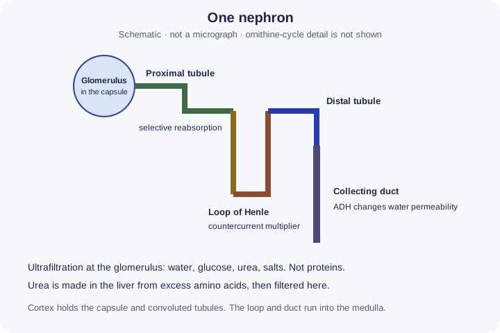
Each kidney has an outer cortex and an inner medulla. Urine collects in the pelvis and leaves through the ureter. The renal arteryA vessel that carries blood away from the heart at high pressure. It has a thick wall with elastic fibres and smooth muscle. brings blood in; the renal veinA vessel that carries blood back to the heart at low pressure. It has a thin wall, a wide lumen and valves. takes filtered blood away. The microscopic unit is the nephronThe functional unit of the kidney, where blood is filtered and urine is formed.. In order: glomerulusA knot of capillaries inside the Bowman’s capsule, where ultrafiltration happens. inside the Bowman’s capsuleThe cup-shaped start of the nephron that surrounds the glomerulus and collects the filtrate., proximal convoluted tubule, loop of HenleThe hairpin loop of the nephron that makes the medulla salty, so water can be reabsorbed from the collecting duct., distal convoluted tubule, and collecting ductThe tube that carries urine through the medulla. ADH controls how much water is reabsorbed from it.. Capsules and convoluted tubules sit mainly in the cortex. The loop and the collecting duct run into the medulla.
Gross structure, then one nephron (continued)
The glomerulus is a knot of capillaryA tiny vessel whose wall is a single layer of endothelium, giving a short diffusion distance for exchange with tissues.. The afferent arteriole is wider than the efferent arteriole, so blood pressureThe pressure that blood exerts on the walls of the blood vessels. in the knot is high. That pressure is the drive for ultrafiltrationFiltration of blood under high pressure in the glomerulus. Small molecules pass into the Bowman’s capsule; cells and large proteins stay behind.. You need both the organ regions and this nephron order.
Urea is made in the liver and filtered in the kidney
Excess amino acidThe monomer of proteins: a central carbon with an amine group, a carboxyl group, a hydrogen and an R group. are not stored as protein. The liver deaminates them: it removes the amino group. Ammonia is toxic, so it is converted to ureaThe main nitrogenous waste product made in the liver from ammonia, and excreted in urine.. The details of the ornithine cycle are not required. The rest of the amino acid, the carbon skeleton, can be respired or converted to carbohydrate or lipid. Urea travels in the blood to the kidney.
At the glomerulus, water, glucoseA hexose (six-carbon) monosaccharide, C₆H₁₂O₆. It is the main respiratory substrate., amino acids, urea and salts are forced into the capsule. The basement membrane holds back proteins and blood cells because they are too large. This is ultrafiltration, a size filter under pressure, not “the kidney chooses urea”. Selection comes later. Urea is not all left behind in the blood; it enters the filtrate.
Proximal tubule, then the loop
The proximal tubuleThe first coiled part of the nephron, where most useful substances are selectively reabsorbed. selectively reabsorbs useful solutes. All of the glucose and amino acids are normally reabsorbed, by active transportMovement of molecules or ions against their concentration gradient through carrier proteins, using energy from ATP. and cotransportTransport of two substances together by one carrier protein, where one moving down its gradient drives the other against its gradient., using ATPAdenosine triphosphate: the molecule that releases energy for cell processes when it is hydrolysed to ADP and Pi.. Some ions are reabsorbed. Water follows by osmosisThe net movement of water molecules from a higher water potential to a lower water potential across a partially permeable membrane.. Microvilli give a large surface areaThe total area of a surface. A larger surface area gives a faster rate of diffusion. and the cells are rich in mitochondrionThe organelle where the link reaction, Krebs cycle and oxidative phosphorylation of aerobic respiration take place.. Glucose in the urine is not the normal result of this specification account. Urea is not reclaimed in the same selective way; it remains to be excreted, although some can move passively.
Proximal tubule, then the loop (continued)
The loop of Henle is a countercurrent multiplierThe way the loop of Henle builds up a high salt concentration deep in the medulla, because the two limbs flow in opposite directions.. The two limbs carry fluid in opposite directions. The ascending limb pumps sodium and chloride ions into the medulla and is impermeable to water, so the medulla becomes concentrated and the fluid in the limb becomes dilute. The descending limb loses water by osmosis into that salty medulla. The countercurrent arrangement builds a gradient along the length of the loop instead of letting one region wash the salts away. A longer loop can support a steeper gradient. Water can then be reabsorbed from the collecting duct if that duct is permeable. Whether it is permeable is the ADHAntidiuretic hormone: released from the posterior pituitary, it makes the collecting duct more permeable to water, so less water is lost in urine. lesson. The loop creates the gradient. It does not, by itself, decide today’s urine concentration.
Follow the process
Excess amino acids are deaminated in the liver → ammonia is converted to urea → urea travels in the blood. At the nephron: wide afferent arteriole and narrower efferent arteriole raise pressure in the glomerulus → water, glucose, amino acids, urea and salts are forced through the basement membrane → proteins and blood cells stay in the blood. Proximal tubule: glucose and amino acids are reabsorbed by active transport, and water follows by osmosis. Loop of Henle: salt is pumped into the medulla and the countercurrent flow builds a gradient. Collecting duct: water leaves only if the duct is permeable. The loop builds the gradient. ADH decides whether the duct uses it. Ornithine-cycle steps are not required.
| Region | Process | What must not be confused |
|---|---|---|
| Glomerulus | Ultrafiltration | Proteins stay in the blood |
| Proximal tubule | selective reabsorptionTaking useful substances, such as glucose, amino acids and much of the water, back into the blood from the filtrate. of glucose and amino acids | Not the urea factory |
| Loop of Henle | Countercurrent multiplier | Not the ADH switch |
| Liver, not a nephron part | Deamination and urea production | Ornithine details are excluded |
Make your reasoning visible
Build the explanation
X happens because Y, leading to Z. X: glucoseA hexose (six-carbon) monosaccharide, C₆H₁₂O₆. It is the main respiratory substrate. is not normally in the urine. Y: it is filtered at the glomerulusA knot of capillaries inside the Bowman’s capsule, where ultrafiltration happens. but then reabsorbed in the proximal tubuleThe first coiled part of the nephron, where most useful substances are selectively reabsorbed. by active transportMovement of molecules or ions against their concentration gradient through carrier proteins, using energy from ATP.. Z: the filtrate that continues has lost its glucose, while ureaThe main nitrogenous waste product made in the liver from ammonia, and excreted in urine., which was made in the liver, is not selectively reclaimed in the same way. Incomplete: “The kidney does not filter glucose.” Improved: “Glucose is filtered, then all of it is normally reabsorbed. Urea is filtered and mostly stays in the fluid that will become urine.”
Liver, then kidney
Urea is produced in the liver. ultrafiltrationFiltration of blood under high pressure in the glomerulus. Small molecules pass into the Bowman’s capsule; cells and large proteins stay behind. removes it from the blood in the kidney.
Multiplier versus switch
The loop builds the salty medulla. ADHAntidiuretic hormone: released from the posterior pituitary, it makes the collecting duct more permeable to water, so less water is lost in urine. decides if the collecting ductThe tube that carries urine through the medulla. ADH controls how much water is reabsorbed from it. uses it.
Avoid this misconception
The claim: The loop of HenleThe hairpin loop of the nephron that makes the medulla salty, so water can be reabsorbed from the collecting duct. filters urea out of the blood, and the ornithine cycle must be drawn.
The correction: Ultrafiltration at the glomerulus puts urea into the filtrate. The loop multiplies a salt gradient for water reabsorption. Ornithine-cycle steps are not required.
How can the kidney make urine more concentrated than your blood?
Water can only move by osmosis, from a higher to a lower water potential. To pull water out of the urine, the kidney first has to make the tissue around the tubule saltier than blood.
What the model shows
One nephron: ultrafiltration at the glomerulus, selective reabsorption in the proximal tubule, the loop of Henle building a salt gradient in the medulla, and ADH controlling how much water the collecting duct reabsorbs.
What to look for
What crosses into the capsule and what stays in the blood. How the medulla gets saltier with each pump and flow. What the urine is like with high and low ADH.
What it tells us
The loop of Henle is a countercurrent multiplier: it builds the gradient. ADH decides whether the collecting duct uses it, so ADH sets the volume and concentration of the urine.
Be careful what you claim
Concentrations are model units, not measured or clinical values. The real loop pumps and flows at the same time, and blood flow in the medulla is left out.
Try it in the model: press Pump step, then Flow step, three times each, and watch the bottom of the medulla.
The nephron and the countercurrent multiplier
Ultrafiltration at the glomerulus, selective reabsorption in the proximal tubule, and a loop of Henle that builds a salt gradient in the medulla. ADH decides whether the collecting duct uses that gradient. Concentrations are model units, not measured values.
What you see
- One nephron across the cortex and medulla: glomerulus and Bowman’s capsule, proximal tubule, loop of Henle, distal tubule and collecting duct.
- The medulla is shaded by the concentration of its tissue fluid, with the value at each level. Orange dots are Na⁺ and Cl⁻ pumped out of the ascending limb; blue drops are water leaving by osmosis.
- On the right, a graph of concentration against depth for the medulla, the ascending limb and the collecting duct, and the filtrate contents for the region you chose.
How to use it
- Click a region of the nephron, or choose it under Region, to see what happens there and what the filtrate contains.
- Press Pump step and Flow step to build the gradient one stage at a time, or Run to repeat them. Change the loop length.
- Move the ADH slider to change how permeable the collecting duct is to water.
What it shows
- The gross and microscopic structure of the kidney and nephron (7.18).
- Urea, made in the liver from excess amino acids, is removed from the blood by ultrafiltration (7.19).
- Selective reabsorption in the proximal tubule, and the loop of Henle as a countercurrent multiplier for water reabsorption (7.20).
- ADH increasing the permeability of the collecting duct, so more water is reabsorbed and less urine is made (7.21).
Limits of the model
- Concentrations are model units, like mOsm per kg but not measured values. They are not clinical thresholds.
- The multiplier is the standard step-by-step teaching model: pump, then flow, with a fixed difference at each level. A real loop pumps and flows at the same time.
- Blood flow in the medulla (the vasa recta) is left out, and volume changes in the loop are ignored. Urine volume is shown as a share of the fluid entering the distal tubule.
- Filtrate contents are shown as all, some or none of what was filtered, not as amounts.
Guided tour · step 1 of 12
- Click the glomerulus in the simulator, or in Nephron, just below these steps, set Region to Glomerulus.
- Read what crosses into the capsule.
High pressure in the glomerulus forces small molecules through the basement membrane. Proteins and blood cells are too large and stay in the blood.
Nephron
Every level starts at the concentration of blood plasma. Press Pump step. Urine about as concentrated as blood. Blood plasma is 300 model units. Urine volume is a share of the fluid entering the distal tubule.
Questions
Check your understanding
Try each question before opening the suggested answer.
Check your understanding (continued)
Practice question
Retrieval and explanation · suggested answer
Name, in order, the five parts of a nephron from glomerulus to duct.
Show suggested answer
Glomerulus in the Bowman’s capsule, proximal convoluted tubule, loop of Henle, distal convoluted tubule, collecting duct.
Practice question
Retrieval and explanation · suggested answer
Where is urea produced, and from what?
Show suggested answer
In the liver, from excess amino acids after deamination. Ornithine-cycle detail is not required.
Practice question
Retrieval and explanation · suggested answer
Why do proteins not enter the filtrate?
Show suggested answer
They are too large to pass the basement membrane during ultrafiltration.
Practice question
Retrieval and explanation · suggested answer
How does the ascending limb help the countercurrent multiplier?
Show suggested answer
It actively transports salts into the medulla and does not let water follow, so the medulla stays concentrated.
Take the idea further
Apply the same specification knowledge to an unfamiliar problem. Any extra assumptions are supplied in the question.
Stretch question
A desert mammal has very long loops of Henle. A student says it therefore never needs ADH. What can the long loop actually explain, and what decision is still left to the collecting duct?
Show a hint
Gradient versus permeability.
Explore the reasoning
A long loop can build a steeper medullary gradient, so more water can be reabsorbed if the collecting duct is permeable. ADH is still what makes that duct permeable. A long loop without permeability would not, on this specification, concentrate the urine by itself. No species fact beyond the question’s premise is required.
Recall this lesson
Lesson recall questions
Question 1 of 5: State where urea is produced from excess amino acids.
Urea and ultrafiltration
Practice question
Retrieval and explanation · suggested answer
State where urea is produced from excess amino acids.
Lesson: 7C 2: Osmoregulation in Mammals: The Kidney
Try this from memory first.
Put your biology to work
Explore where these ideas appear in the world. Enrichment, not extra required recall.
Filter and reabsorb, then the specification adds the mechanism
7C · Osmoregulation in Mammals: The KidneyNIDDK describes kidneys as filtering blood, removing wastes such as urea and extra water, and returning water and nutrients the body still needs. It names the nephron, the…Specification 7.20Read & use the scienceClose article
Why this matters
NIDDK describes kidneys as filtering blood, removing wastes such as urea and extra water, and returning water and nutrients the body still needs. It names the nephron, the glomerulus and the tubule. That is the right public outline. It does not teach the afferent arteriole, the basement membrane, or the loop as a countercurrent multiplier. Those mechanisms stay in 7.18–7.20. 7.18–7.20: structure, urea and ultrafiltration, selective reabsorption, and the loop of Henle.
Back to the specification
Pearson Edexcel IAL Biology · Issue 2, February 2021 · Unit 5, pp. 35–38. The application extends beyond required recall.
- 7.20 Selective reabsorption and the loop of Henle.
Use the science
Why is “the tubule filters like a sieve” less precise than the specification?
Explore a possible response
The sieve-like pressure filter is the glomerulus. The tubule then reabsorbs useful solutes. Calling the whole tubule a sieve misses selective reabsorption and the multiplier.
Explore the source
- NIDDK: your kidneys and how they workUS National Institute of Diabetes and Digestive and Kidney Diseases
A dialyser is selective in a different way
7C · Osmoregulation in Mammals: The KidneyThe NIDDK describes haemodialysis as blood flowing through a filter while dialysis fluid moves on the other side. Waste solutes and excess water can leave the blood, but…Specification 7.20Read & use the scienceClose article
Why this matters
The NIDDK describes haemodialysis as blood flowing through a filter while dialysis fluid moves on the other side. Waste solutes and excess water can leave the blood, but the treatment replaces only part of kidney function. A nephron filters at the glomerulus and then selectively reabsorbs useful substances along its tubule; the loop of Henle helps build the medullary gradient for water conservation. A dialyser does not reconstruct that living countercurrent system. A renal specialist must adjust dialysis fluid and treatment conditions using measurements, which is why a “machine kidney” is an analogy with important limits.
Back to the specification
Pearson Edexcel IAL Biology · Issue 2, February 2021 · Unit 5, pp. 35–38. The application extends beyond required recall.
- 7.20 Selective reabsorption and the loop of Henle.
Use the science
Why would a dialyser that removes urea not automatically reproduce selective reabsorption in a healthy proximal tubule?
Explore a possible response
Removing a waste solute through a membrane is different from active, selective recovery of useful solutes into blood. The machine lacks the nephron’s living transport cells and countercurrent organisation.
Explore the source
- NIDDK: haemodialysisUS National Institute of Diabetes and Digestive and Kidney Diseases