
What you need to know
- 6.18gel electrophoresisSeparating DNA fragments by size: they move through a gel towards the positive electrode, and smaller fragments move further. separates DNADeoxyribonucleic acid: a double-stranded polynucleotide that carries the genetic code. fragments of different lengths.
- 6.19DNA profilingComparing the lengths of non-coding repeated DNA sequences to identify individuals or show relationships. identifies organisms and shows genetic relationships in plants and animals.
A teaching gel is not a courtroom statistic. One locus is not an identification. Identical twins share a profile. Profiling is not the same as sequencing every gene. The bands in the figure are fictional.
By the end, you should be able to…
- Explain why shorter DNA fragments travel further.
- Describe a profile as a pattern of repeat-length alleleA different version of the same gene, found at the same locus..
- Use a parent-and-child pattern without claiming certainty from one locusThe position of a gene on a chromosome..
Build your understanding
The gel sorts by length
DNADeoxyribonucleic acid: a double-stranded polynucleotide that carries the genetic code. fragments are loaded into a well in a gel, usually agarose, covered with buffer. A direct current is applied. Phosphate groups make DNA negatively charged, so fragments move toward the positive electrode, the anode. The gel is a mesh. Shorter fragments thread through faster and travel further in the same time. Longer fragments lag behind.
A lane of known lengths, a size ladder, lets you estimate how long an unknown band is. A stain makes the DNA visible afterwards. Some stains are hazardous, so a school uses the dye it has assessed. Electrophoresis separates lengths. It does not read the base sequence.
A profile is a pattern of lengths
DNA profilingComparing the lengths of non-coding repeated DNA sequences to identify individuals or show relationships. uses regions where a short sequence is repeated. Different people, or different individuals of a plant or animal speciesA group of organisms that can interbreed to produce fertile offspring., can have different numbers of repeats. polymerase chain reactionA method that makes many copies of a DNA sequence by repeated cycles of heating (denaturing), cooling (annealing primers) and extending with Taq polymerase. copies those regions. Electrophoresis separates the copies. The positions of the bands are the alleleA different version of the same gene, found at the same locus.. A profile is the pattern across several such regions, not one band.
The specification includes plants and animals, not only humans. The same idea identifies a cultivar, a poached animal, or a person. Non-coding repeats are chosen so that the test is not reading a disease geneA sequence of DNA bases that codes for a polypeptide or a functional RNA.. You do not need a list of commercial locusThe position of a gene on a chromosome. names.
Relationships, and the limits
At one locus a child inherits one allele from each parent, so each of the child’s bands should match one parent or the other. A band that matches neither parent needs an explanation: mutationA change in the sequence of bases in DNA. is rare, a laboratory error or a wrong assumed parent is more likely in a teaching question. Across many loci, a matching pattern is strong evidence of identity. Identical twins have the same DNA, so a profile does not tell them apart.
A partial profile, a mixed sample, or contamination can look like a match. NIST’s forensic work on difficult DNA samples is a reminder that the laboratory method matters. Do not invent a probability such as “one in a billion” unless the question gives it. Do not say the gel proves guilt. It is identification evidence.
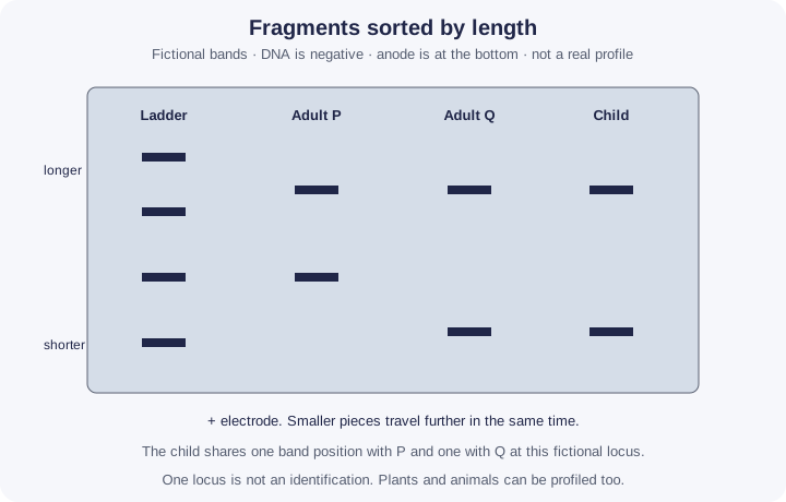
| Observation | Reason | What it is not |
|---|---|---|
| Band further down | Shorter fragment | Not “more DNA” by itself |
| Two bands in one lane | Two alleles of different length | Not two unrelated people, unless the lane was mixed |
| Child matches one band from each adult | Possible inheritance at that locus | Not proof from one locus alone |
Make your reasoning visible
Charge, then mesh
Negative DNADeoxyribonucleic acid: a double-stranded polynucleotide that carries the genetic code. moves to the positive electrode. The mesh makes short pieces go further.
Many loci
Say that identification uses the combination. One band is a length, not a name.
Avoid this misconception
The claim: The furthest band is the largest fragment, and a matching profile is unique even for identical twins.
The correction: The furthest band is the shortest fragment. Identical twins share the same profile. A match is strong evidence when many loci agree, not a magical certainty from one lane.
How can a gel tell two people apart?
People differ in how many times short sequences are repeated in some non-coding regions of DNA. PCR copies those regions; a gel sorts the copies by length.
What the model shows
Gel electrophoresis of fictional repeat regions beside a size ladder, for a crime scene and for a family.
What to look for
Which way the DNA moves and which bands travel furthest. How many suspects match with one locus, and with two.
What it tells us
Shorter fragments travel further. A profile is the pattern of lengths across several loci, and a child shares one band with each parent at each locus.
Be careful what you claim
All bands are fictional and there are only two loci. Real profiles use many loci; a match is evidence, not proof.
Try it in the model: press Run the gel, then Stain the gel, and find the band that travelled furthest.
Gel electrophoresis and DNA profiles
PCR copies a chosen region of DNA, doubling it each cycle in theory. A gel then sorts the copies by length beside a size ladder. All copy numbers are model values and all samples are fictional.
What you see
- PCR: a thermocycler trace for one cycle (95, 55 and 72 °C) and one DNA molecule going through it: the strands separate, the primers bind and Taq builds new strands.
- A log graph of target copies against cycle number: the theoretical 2ⁿ line and a real tube that levels off.
- Gel: a size ladder and four fictional samples. The loading dye moves while the gel runs; the bands appear when you stain it. A standard curve of distance against length sits beside it.
How to use it
- Under Part, choose PCR or Gel.
- PCR: Next step moves one stage at a time. Run 1 cycle animates a cycle. Fast-forward runs 5 or to 30 cycles. Change the starting molecules and the sample.
- Gel: set the voltage and press Run the gel, then Stain the gel. Click a band to estimate its length. Change the number of loci and the sample set.
What it shows
- DNA amplified by the polymerase chain reaction (6.17): the three temperatures, primers, Taq and the 2ⁿ calculation.
- Gel electrophoresis separating DNA fragments by length (6.18).
- DNA profiling to identify individuals and show relationships, in people, plants and animals (6.19).
Limits of the model
- Copy numbers are model values. The real tube is a simple model of efficiency falling as reagents run out.
- Strictly, fragments of exactly the target length first appear in cycle 3, so the exact count is 2ⁿ − 2n from one molecule. The exam uses starting number × 2ⁿ, as this model does.
- All band sizes are fictional and there are only two loci. Real profiles use many loci, and a match is evidence, not a probability or proof of guilt.
- Distance is modelled as falling with log(length) over the gel’s working range; real gels depend on the agarose percentage and buffer.
Guided tour · step 2 of 8
- In Gel, press Run the gel.
- Watch the blue loading dye move toward the positive electrode.
Phosphate groups make DNA negatively charged, so the fragments move toward the anode (+). The dye shows how far the run has gone.
Gel
Click a band to read its length against the ladder. Higher voltage runs the gel faster; the dye front stops the run before small fragments run off the end.
Questions
Check your understanding
Try each question before opening the suggested answer.
Check your understanding (continued)
Practice question
Retrieval and explanation · suggested answer
Why does DNA move toward the anode?
Show suggested answer
DNA is negatively charged because of its phosphate groups, and the anode is the positive electrode.
Practice question
Retrieval and explanation · suggested answer
Two fragments are 200 and 800 base pairs. Which travels further in the same time, and why?
Show suggested answer
The 200-base-pair fragment. Shorter pieces move more easily through the gel.
Practice question
Retrieval and explanation · suggested answer
A child has band positions that are absent from both proposed parents at several loci. What is the cautious conclusion?
Show suggested answer
The child is unlikely to be the offspring of that pair, or the samples were mixed up. One odd locus could be an error. Several loci make a relationship unlikely.
Practice question
Retrieval and explanation · suggested answer
Why can the same method be used on a plant?
Show suggested answer
Plants also have DNA regions that differ in length between individuals. Profiling is not limited to humans.
Take the idea further
Apply the same specification knowledge to an unfamiliar problem. Any extra assumptions are supplied in the question.
Stretch question
Fictional gel, one locus: adult P has bands at 200 and 400 bp, adult Q at 200 and 600 bp, young plant at 400 and 600 bp. Could the young plant be the offspring of P and Q? A second locus in the young plant matches neither adult. What changes?
Show a hint
Check one locus, then do not ignore the second.
Explore the reasoning
At the first locus the young plant could have inherited 400 from P and 600 from Q. The shared 200 is not required in the offspring. The second locus, matching neither adult, means this pair is unlikely to be the two parents unless that lane failed. One supporting locus does not outweigh a clear mismatch. Lengths are invented.
Recall this lesson
Lesson recall questions
Question 1 of 5: How does an electric field separate DNA fragments in agarose gel?
Gel electrophoresis
Practice question
Retrieval and explanation · suggested answer
How does an electric field separate DNA fragments in agarose gel?
Try this from memory first.
Put your biology to work
Explore where these ideas appear in the world. Enrichment, not extra required recall.
Length, not a name printed on the band
6C · DNA ProfilingYourgenome describes gel electrophoresis as using a current to separate charged DNA by size: DNA is negative, it moves toward the positive electrode, and shorter fragments travel further.…Specification 6.18Read & use the scienceClose article
Why this matters
Yourgenome describes gel electrophoresis as using a current to separate charged DNA by size: DNA is negative, it moves toward the positive electrode, and shorter fragments travel further. A size marker lets you judge length. The page is about the separation. It does not turn a band into a person’s name. Profiling is what you do with several such lengths. The size marker and several loci make a profile interpretable. A single band shared by two lanes is weak evidence because unrelated organisms can share fragment lengths at one locus.
Back to the specification
Pearson Edexcel IAL Biology · Issue 2, February 2021 · Unit 4, pp. 29–32. The application extends beyond required recall.
- 6.18 know how gel electrophoresis can be used to separate DNA fragments of different length
Use the science
Why is “the band travelled furthest, so it is the most important gene” a misunderstanding of the gel?
Explore a possible response
Distance measures length, not importance. In a profile the fragments are usually non-coding repeats. The furthest band is simply the shortest piece in that lane.
Explore the source
- Yourgenome: what is gel electrophoresis?Wellcome Genome Campus
A DNA band is not a verdict
6C · DNA ProfilingNIST explains why a forensic DNA mixture is harder to interpret than a clean textbook gel. Several people may contribute DNA, a small amount can produce weak signals,…Specification 6.18 · 6.19Read & use the scienceClose article
Why this matters
NIST explains why a forensic DNA mixture is harder to interpret than a clean textbook gel. Several people may contribute DNA, a small amount can produce weak signals, and some alleles can drop out during testing. Electrophoresis still separates fragments by length; interpretation then compares several genetic markers and considers uncertainty. A crime-drama image of one matching band cannot identify a person by itself. The analyst must also ask whether the sample is relevant, whether contamination is possible and how strong the comparison is. A profile can support an inference about identity or relationship, not a direct conclusion about what action occurred.
Back to the specification
Pearson Edexcel IAL Biology · Issue 2, February 2021 · Unit 4, pp. 29–32. The application extends beyond required recall.
- 6.18 know how gel electrophoresis can be used to separate DNA fragments of different length
- 6.19 understand how DNA profiling is used for identification and determining genetic relationships between organisms (plants and animals)
Use the science
Why is one band of matching length much weaker evidence than a well-controlled multi-locus profile?
Explore a possible response
Many people can share one fragment length. Multiple independent markers and controls reduce coincidence and reveal mixture or contamination issues.