
What you need to know
- 6.9antigenA molecule, often a protein or glycoprotein on a cell surface, that the immune system recognises as non-self. and antibodyA Y-shaped protein made by plasma cells that binds specifically to one antigen.; plasma cellA cell formed from a B effector cell that secretes large amounts of one antibody., macrophages and antigen-presenting cellA cell, such as a macrophage, that displays a pathogen’s antigens on its surface so that T helper cells can bind..
- 6.10B memory cellA long-lived B cell that stays after an infection and quickly divides into plasma cells if the same antigen returns. and B effector cellAn activated B cell from the clone that differentiates into a plasma cell to secrete antibodies.; T helper cellA T cell that binds to an antigen-presenting cell and releases cytokines that activate B cells and T killer cells., T killer cellA T cell that binds to infected body cells showing the matching antigen and destroys them. and T memory cellA long-lived T cell that stays after an infection, allowing a faster secondary response..
Non-specific processes stay in 6B 1. Types of immunity (vaccine versus infection) are 6B 3. HIV killing T helpers is used as a 6.10 application, not a repeat of the 6A case study.
By the end, you should be able to…
- Define antigen and antibody, and use the Y-shaped variable regionThe part of an antibody that differs between antibodies and forms the antigen-binding site..
- State roles of plasma cells and antigen-presenting macrophages.
- Distinguish B effector, B memory, T helper, T killer and T memory.
Build your understanding
Antigen in, antibody out
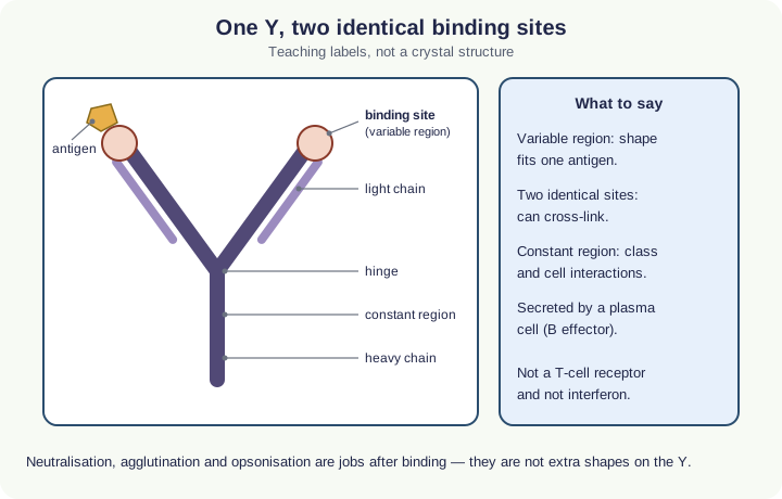
An antigen is a molecule, often a protein, that the immune system recognises as non-self and that can trigger a response. An antibodyA Y-shaped protein made by plasma cells that binds specifically to one antigen. is a specific immunoglobulinAnother name for an antibody. secreted by a plasma cellA cell formed from a B effector cell that secretes large amounts of one antibody.. The Y has two identical variable regionThe part of an antibody that differs between antibodies and forms the antigen-binding site. that form antigen-binding sites complementary to one antigen, so one antibody can cross-link two identical antigenA molecule, often a protein or glycoprotein on a cell surface, that the immune system recognises as non-self.. The constant regionThe part of an antibody that is the same in all antibodies of one class. Phagocytes can bind to it. sets the class and how the molecule interacts with other cells.
After binding, antibodies can neutralise toxins or binding sites, agglutinate pathogens, and opsonise them so phagocyteA white blood cell, such as a neutrophil or macrophage, that engulfs and digests pathogens. engulf them more readily. Those are jobs of bound antibody, not extra 6.8 processes.
Who shows the antigen, who secretes
A macrophage that has phagocytosed a pathogen can act as an antigen-presenting cellA cell, such as a macrophage, that displays a pathogen’s antigens on its surface so that T helper cells can bind. (APC): it displays processed antigen on its surface. A T helper cellA T cell that binds to an antigen-presenting cell and releases cytokines that activate B cells and T killer cells. with a matching receptor binds that displayed antigen, is activated, and secretes cytokineA signalling protein released by immune cells, such as T helper cells, that stimulates other immune cells.. MHC proteins that hold the peptide are enrichment naming, not extra 6.9 recall. That is why 6.8 phagocytosisEngulfing a pathogen or particle into a vesicle by endocytosis. and 6.9 presentation are linked but not the same sentence.
clonal selectionSelection of the lymphocyte whose receptors are complementary to an antigen. It then divides by mitosis to form a clone. activates only the B or T clone whose receptor fits the antigen. B effector cellAn activated B cell from the clone that differentiates into a plasma cell to secrete antibodies. (plasma cells) then secrete large amounts of that one antibody. Each plasma-cell clone has one specificityAn enzyme catalyses only one reaction because only one substrate has a shape complementary to its active site. because it came from one selected B cell.
Follow the process
A macrophage engulfs the pathogen and displays antigen as an antigen-presenting cell → a matching T helper binds and secretes cytokines → only the B or T clone whose receptor fits that antigen is selected → B effector cells (plasma cells) secrete antibody with a variable region complementary to it → B memory cellA long-lived B cell that stays after an infection and quickly divides into plasma cells if the same antigen returns. remain, so a later meeting gives a faster, larger response. T killer cellA T cell that binds to infected body cells showing the matching antigen and destroys them. destroy infected host cells. They do not secrete the antibody.
Five named lymphocytes
B effector cells secrete antibody. B memory cells remain so a later meeting with the same antigen gives a faster, larger antibody response. T helper cells secrete cytokines that activate B cells, T killers and macrophages. T killer (cytotoxic T) cells destroy infected host cells, for example by releasing perforinA protein released by T killer cells that makes holes in the membrane of an infected cell, killing it.. T memory cellA long-lived T cell that stays after an infection, allowing a faster secondary response. speed a later cellular response.
Loss of T helpers, as in untreated HIVHuman immunodeficiency virus: an RNA retrovirus that infects T helper cells and can lead to AIDS. (6A 8), removes the cytokines both antibody and T-killer paths need, so both collapse. Neutrophils are phagocytes, not a type of T cell. Do not list every immunoglobulin class unless a question gives it.
NOT: T killer cells do not secrete antibodies. Macrophages are not B cells.
- An antibody has two identical antigen-binding sites
- Bound antibody can neutralise, agglutinate or opsonise
- T memory cells speed a later cellular response
- Loss of T helpers, as in untreated HIV, collapses both paths
- MHC proteins are enrichment naming, not required recall
Two traps this diagram blocks
- Do not say T killer cells secrete antibodies.
- Do not say every lymphocyte responds to every antigen.
From the diagram alone
Question
Explain the roles of T helper cells and plasma cells in producing antibody. [4]
Show a model answer
A T helper cell with a receptor that fits the displayed antigen is activated and secretes cytokines. Those cytokines help activate the matching B clone. The B effector cells, plasma cells, then secrete antibody with a binding site complementary to that antigen.
| Cell | Lineage | Job |
|---|---|---|
| B effector / plasmaThe liquid part of blood: water carrying dissolved substances such as glucose, ions, hormones and proteins. | B | Secretes specific antibody |
| B memory | B | Faster secondary antibody response |
| T helper | T | Cytokines that activate others |
| T killer | T | Destroys infected host cells |
| T memory | T | Faster secondary cellular response |
Make your reasoning visible
Build the explanation
X happens because Y, leading to Z. X: a large amount of one antibody appears. Y: clonal selectionSelection of the lymphocyte whose receptors are complementary to an antigen. It then divides by mitosis to form a clone. activates the B cell whose receptor matches the antigenA molecule, often a protein or glycoprotein on a cell surface, that the immune system recognises as non-self., and cytokineA signalling protein released by immune cells, such as T helper cells, that stimulates other immune cells. from a T helper cellA T cell that binds to an antigen-presenting cell and releases cytokines that activate B cells and T killer cells. help that clone become plasma cellA cell formed from a B effector cell that secretes large amounts of one antibody.. Z: the variable regionThe part of an antibody that differs between antibodies and forms the antigen-binding site. fits that antigen, and memory cells remain. Incomplete: “The body makes antibodyA Y-shaped protein made by plasma cells that binds specifically to one antigen..” Improved: “An antigen-presenting cellA cell, such as a macrophage, that displays a pathogen’s antigens on its surface so that T helper cells can bind. macrophage activates a matching T helper, which releases cytokines so the selected B clone secretes antibody to that antigen.”
Variable region = specificity
The stem (constant regionThe part of an antibody that is the same in all antibodies of one class. Phagocytes can bind to it.) does not choose the antigen.
Name cytokines for T helpers
“They help” is not a mechanism.
Avoid this misconception
The claim: T killer cellA T cell that binds to infected body cells showing the matching antigen and destroys them. secrete antibodies, and macrophages are a kind of B cell.
The correction: Antibodies come from B effectors (plasma cells). Macrophages phagocytose and can present antigen. T killers destroy infected host cells.
Why is the second infection with the same pathogen so much milder?
The body first responds to any pathogen in the same way. Then, over several days, it builds a response aimed at one antigen and keeps cells that remember it.
What the model shows
A bacterium or a virus going from the non-specific opening, through antigen presentation and clonal selection, to antibody, T killer cells and memory.
What to look for
Which steps need a receptor that fits the antigen. How long the antigen takes to produce antibody the first time and the second time.
What it tells us
Only lymphocytes with complementary receptors are selected and cloned. Memory cells make the secondary response faster, larger and longer-lasting.
Be careful what you claim
The times and antibody levels are model numbers, and the steps overlap more in a real infection.
Try it in the model: press Next step until the B cells appear, then click a B cell whose receptor does not fit and read why it is not selected.
From phagocytosis to the specific response
Choose a bacterium or a virus and step through the response. The opening steps are non-specific. Cytokines and antibody appear only after a matching lymphocyte is selected. The drawings are a model, and the days and antibody levels are model values, not measured data.
What you see
- A timeline of the response across the top: the non-specific steps, then the specific response from antigen presentation onwards.
- A drawing of the current step, with the cells and chemical signals active at that step listed underneath.
- A graph of antibody concentration in the blood against time, in model units.
How to use it
- Choose a bacterium or a virus, then press Next step. Click a visited step on the timeline to go back to it.
- At clonal selection, click the lymphocyte whose receptor fits the antigen, or pick its number under the model.
- After the memory step, press Meet the same antigen again to see the secondary response.
What it shows
- Inflammation, lysozyme, interferon and phagocytosis are non-specific responses (6.8).
- Macrophages act as antigen-presenting cells. T helper cytokines activate the matching B cells, which become plasma cells that secrete antibody (6.9, 6.10).
- T killer cells destroy infected cells, and B and T memory cells give a faster, larger secondary response (6.10, 6.11).
Limits of the model
- The days and antibody levels are model values, not measured data. Real lags and peaks vary.
- Receptor and antigen shapes are symbols for complementary shapes; real receptors bind a small part of an antigen.
- Each step is shown one at a time. In the body many of them overlap.
Guided tour · step 1 of 10
- In Response, just below these steps, keep Pathogen on Bacterium (options: Bacterium, Virus).
- Press Next step through inflammation, the phagosome and the phagolysosome, as far as the T helper.
Inflammation, lysozyme and phagocytosis act on any pathogen. The macrophage then displays the antigen and becomes an antigen-presenting cell.
Response
Histamine, lysozyme and interferon are non-specific. Cytokines come from T helpers. Antibody comes from plasma cells. A lysosome is the organelle in the phagocyte; lysozyme is the enzyme that hydrolyses bacterial peptidoglycan.
Questions
Final check: put it in order
What to do
- Pick a chain with Chain, just below these steps: Humoral (B cells) or Cell-mediated (T killer).
- Reorder the cards below: drag a card (on a touch screen, drag its number), tap a card then tap where it goes, or use the ↑ and ↓ arrows at its right.
- Press Check under the cards to mark each one green (right place) or red; the count shows how many are right.
- Stuck? Hint first outlines the first wrong place, then gives a clue, then places that step for you.
- Shuffle mixes the cards again; Start again returns them to the starting order. Keyboard: Space picks up, arrow keys move, Space drops.
A bacterium has entered the blood. Put the humoral response in order.
0 of 8 in the right place
Antigen presentation and the activation of T helpers are simplified. Memory cells are made during clonal expansion; they are listed last because they last after the infection.
Check your understanding
Try each question before opening the suggested answer.
Check your understanding (continued)
Practice question
Retrieval and explanation · suggested answer
Define antigen and antibody.
Show suggested answer
An antigen is a non-self molecule that can trigger an immune response. An antibody is a specific immunoglobulin secreted by a plasma cell that binds that antigen at its variable region.
Practice question
Retrieval and explanation · suggested answer
State the role of a macrophage as an antigen-presenting cell.
Show suggested answer
It phagocytoses the pathogen and displays processed antigen on its surface so a matching T helper can recognise it.
Practice question
Retrieval and explanation · suggested answer
List the two B-cell types and three T-cell types named in 6.10.
Show suggested answer
B memory and B effector; T helper, T killer and T memory.
Practice question
Retrieval and explanation · suggested answer
Why does loss of T helper cells undermine both antibody and T-killer responses?
Show suggested answer
T helper cytokines are needed to activate B cells and T killers, so without them fewer plasma cells and T killers are produced.
Take the idea further
Apply the same specification knowledge to an unfamiliar problem. Any extra assumptions are supplied in the question.
Stretch question
Using the teaching Y, explain why one plasma-cell clone cannot secrete antibodies against both TMV and HIV, then say which 6.10 cell would still be useful against an HIV-infected T helper.
Show a hint
One variable region; infected host cell.
Explore the reasoning
Clonal selection gives each plasma cell one complementary variable region, so its antibody fits one antigen, not both viruses. An HIV-infected T helper is an infected host cell, so T killers (with T helper cytokines, which HIV itself destroys) are the 6.10 cell that targets infected cells. Antibody against free virus is a different job.
Recall this lesson
Lesson recall questions
Question 1 of 5: State the role of plasma cells in the immune response.
Antigens and antibodies
Practice question
Retrieval and explanation · suggested answer
State the role of plasma cells in the immune response.
Lesson: 6B 2: The Specific Response to Infection
Try this from memory first.
Put your biology to work
Explore where these ideas appear in the world. Enrichment, not extra required recall.
NIAID’s adaptive layer is clonal and specific
6B · The Specific Response to InfectionNIAID describes adaptive immunity as lymphocyte responses that recognise particular antigens and can remember them. That is 6.9–6.10 in other clothes: APC, matching receptor, clone, antibody or T…Specification 6.9Read & use the scienceClose article
Why this matters
NIAID describes adaptive immunity as lymphocyte responses that recognise particular antigens and can remember them. That is 6.9–6.10 in other clothes: APC, matching receptor, clone, antibody or T killer, memory. Their extra named subsets are enrichment. Required names stay plasma/B effector, B memory, T helper, T killer, T memory. Specificity comes from matching receptors and antigens, not from every B or T cell responding equally. The first encounter expands a suitable clone; memory cells can then support a faster later response.
Back to the specification
Pearson Edexcel IAL Biology · Issue 2, February 2021 · Unit 4, pp. 29–32. The application extends beyond required recall.
- 6.9 understand the roles of antigens and antibodies in the body’s immune response including the involvement of plasma cells, macrophages and antigen-presenting cells
Use the science
Why is “the macrophage secreted the antibody after it ate the bacterium” a scrambled 6.9 answer?
Explore a possible response
The macrophage presents antigen. The plasma cell secretes antibody. Presentation and secretion are different cells.
Explore the source
An antibody becomes a cancer medicine
6B · The Specific Response to InfectionThe US National Cancer Institute explains that laboratory-made monoclonal antibodies can bind chosen proteins on cancer cells. Some mark cells for immune attack; others bring T cells near…Specification 6.9 · 6.10Read & use the scienceClose article
Why this matters
The US National Cancer Institute explains that laboratory-made monoclonal antibodies can bind chosen proteins on cancer cells. Some mark cells for immune attack; others bring T cells near a target. This uses the specificity familiar from an antibody–antigen diagram, but cancer therapy adds careful target selection and clinical testing. A marker is not automatically unique to cancer, so treatment may also affect healthy cells carrying it. Natural plasma cells secrete antibodies after B-cell activation; a manufactured therapeutic antibody is supplied from outside the patient. That distinction prevents the mistaken claim that an infused antibody itself creates B-memory cells.
Back to the specification
Pearson Edexcel IAL Biology · Issue 2, February 2021 · Unit 4, pp. 29–32. The application extends beyond required recall.
- 6.9 understand the roles of antigens and antibodies in the body’s immune response including the involvement of plasma cells, macrophages and antigen-presenting cells
- 6.10 understand the differences between the roles of B cells (B memory and B effector cells), and T cells (T helper, T killer and T memory cells) in the host’s immune response
Use the science
Why might a monoclonal antibody against a protein also affect healthy cells?
Explore a possible response
If healthy cells express the same target protein, the antibody can bind them too; antigen specificity is not the same as cancer-only expression.
Explore the source
- Monoclonal antibodies in cancer treatmentUS National Cancer Institute