
What you need to know
- 4.5sclerenchymaPlant tissue of dead cells with thick lignified walls, such as fibres, that gives support. fibres, xylemPlant tissue that carries water and mineral ions up from the roots. Its vessels are dead, hollow and lignified. vessels and phloemPlant tissue that transports sucrose and other organic substances, in sieve tubes with companion cells.: structure, stem position and function.
- 4.6Core Practical 7: plan diagramA drawing that shows the outlines of tissues and where they are, with no individual cells drawn. and identification of these tissues.
Specification 4.5 names sclerenchyma fibres, xylem vessels and phloem. Collenchyma may appear in textbooks as living support tissue with uneven cellulose thickening; it is not one of the three named tissues. Companion cells support phloem function but are not a fourth named 4.5 tissue.
By the end, you should be able to…
- Compare xylem, phloem and sclerenchyma fibres by structure and function.
- State their positions in a typical dicot stem.
- Distinguish a plan diagram from a high-power cell drawing.
Build your understanding
Three tissues, three jobs
xylemPlant tissue that carries water and mineral ions up from the roots. Its vessels are dead, hollow and lignified. vessels are dead, hollow tubes with lignifiedDescribes a cell wall strengthened and waterproofed by lignin. walls. End walls are lost or heavily perforated, and pits allow lateral water movement. They transport water and mineral ions and provide support. ligninA tough, waterproof polymer laid down in the walls of xylem vessels and sclerenchyma fibres. waterproofs the wall and stops it collapsing when water is pulled under tension.
phloemPlant tissue that transports sucrose and other organic substances, in sieve tubes with companion cells. translocates organic solutes such as sucroseA disaccharide of glucose and fructose. It is the sugar transported in phloem.. Sieve-tube elements are living, with sieve plates and reduced contents. companion cellA phloem cell next to a sieve tube element. It has many mitochondria and supports the sieve tube. are nucleated and metabolically active; they load solutes into the sieve tubeA phloem tube of living cells with few organelles, joined end to end through sieve plates.. Phloem is not lignified like xylem and is not a mass-flow water pipe from root to leaf.
sclerenchymaPlant tissue of dead cells with thick lignified walls, such as fibres, that gives support. fibres are long, tapering cells with thick lignified secondary walls. They are dead at maturity and provide support. They do not transport water as xylem vessels do, and they are not the living translocation pathway.
Where to look in a stem
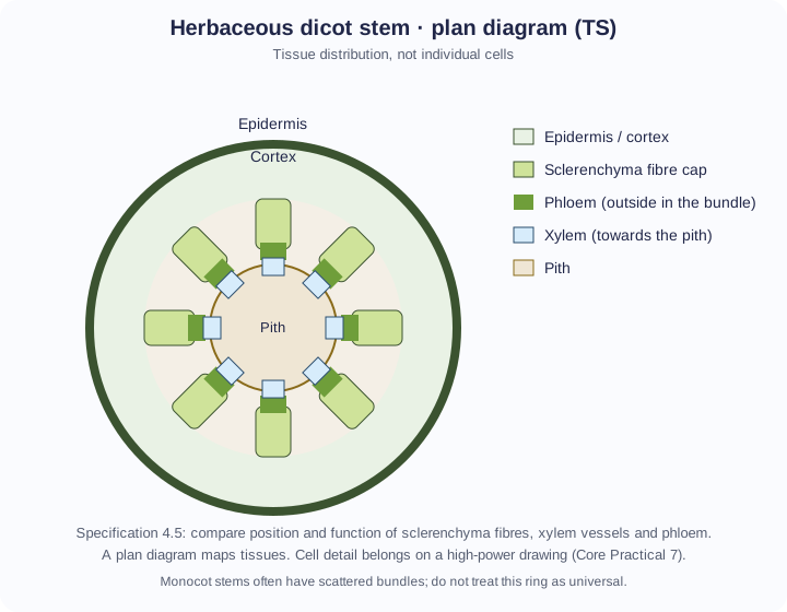
In a typical herbaceous dicot stem, vascular bundles form a ring around the pith. Within a bundle, xylem lies towards the inside (the pith) and phloem towards the outside. Sclerenchyma fibres often form a cap outside the phloem. Epidermis and cortex lie outside the ring; pith occupies the centre.
Where to look in a stem (continued)
Roots and leaves arrange the same tissues differently. In a typical dicot leaf veinA vessel that carries blood back to the heart at low pressure. It has a thin wall, a wide lumen and valves., xylem is towards the upper side and phloem towards the lower side. In a typical dicot root, xylem may occupy a central star with phloem between the arms. Always state the organ you are describing.
Plan first, then cells
A plan diagramA drawing that shows the outlines of tissues and where they are, with no individual cells drawn. shows tissue distribution with clear outlines and no individual cells or shading. A high-power drawing shows cell shapes, wall thickness and contents you can actually see. Mixing the two is a common practical error.
Core Practical 7 asks you to draw both kinds of record and to identify sclerenchyma fibres, phloem sieve tubes and xylem vessels with their locations. Labels must match visible evidence: thick lignified walls and empty lumens for xylem; thin-walled living sieve tubes with companion cells outside the xylem; very thick-walled fibres with tiny lumens in a cap outside the phloem.
Follow the process
Water in xylem: a continuous column in dead, hollow vessels (end walls lost, no cytoplasmEverything inside the cell surface membrane except the nucleus: the fluid cytosol and the organelles. to obstruct flow) → cohesionThe attraction of water molecules to one another through hydrogen bonds. between water molecules lets tension pull the column → lignin stops the walls collapsing under that tension → pits in non-lignified regions let water move sideways if one route is blocked. Small diameter also helps the column hold together. This is transport under tension, not a pump inside the vessel.
Sucrose in phloem is a different line. Companion cells use ATPAdenosine triphosphate: the molecule that releases energy for cell processes when it is hydrolysed to ADP and Pi. to load sucrose into a sieve tube at a source → the soluteA substance that is dissolved in a solvent. lowers water potentialThe tendency of water to move out of a solution. Pure water is 0; adding solutes makes it negative. Water moves from higher to lower water potential. → water enters from xylem → pressure pushes sap through sieve plates towards a sink → sucrose is unloaded. Sieve tubes stay living but have little cytoplasm, which leaves space for flow. Do not describe phloem as a lignified, dead water pipe.
| Tissue | Living at maturity? | Main function | Typical stem position |
|---|---|---|---|
| Xylem vessels | No | Water and mineral transport; support | Inside of the vascular bundle, towards the pith |
| Phloem (sieve tubes) | Yes | Translocation of organic solutes | Outside of the bundle, towards the epidermis |
| Sclerenchyma fibres | No | Support | Often a cap outside the phloem |
Make your reasoning visible
Build the explanation
X happens because Y, leading to Z. X: the water column keeps moving through a xylemPlant tissue that carries water and mineral ions up from the roots. Its vessels are dead, hollow and lignified. vessel. Y: there is no cytoplasmEverything inside the cell surface membrane except the nucleus: the fluid cytosol and the organelles. or end wall to block it, and cohesionThe attraction of water molecules to one another through hydrogen bonds. lets tension pull the molecules together. Z: ligninA tough, waterproof polymer laid down in the walls of xylem vessels and sclerenchyma fibres. keeps the tube from collapsing. Incomplete: “Xylem is dead so it transports water.” Improved: “Mature vessels are hollow and lignifiedDescribes a cell wall strengthened and waterproofed by lignin., so water can move under tension without the wall collapsing.” The improvement links the structure to the force. For phloemPlant tissue that transports sucrose and other organic substances, in sieve tubes with companion cells., name companion-cell loading and pressure, not lignin.
Name the organ
Bundle position in a dicot stem is not automatically the same in a root or leaf.
Avoid this misconception
The claim: Xylem and phloem are both dead pipes that carry water.
The correction: Xylem vessels are dead tubes for water and mineral ions. Phloem sieve tubeA phloem tube of living cells with few organelles, joined end to end through sieve plates. are living and translocate organic solutes.
Check your understanding
Try each question before opening the suggested answer.
Practice question
Retrieval and explanation · suggested answer
Compare xylem vessels and sclerenchyma fibres as supporting tissues.
Show suggested answer
Both have lignified secondary walls and are dead at maturity, giving strength. Xylem vessels are hollow tubes that also transport water and mineral ions; sclerenchyma fibres are long support cells without that transport role.
Practice question
Retrieval and explanation · suggested answer
In a typical dicot stem TS, where is phloem relative to xylem in a vascular bundle?
Show suggested answer
Phloem is towards the outside of the bundle; xylem is towards the pith.
Practice question
Retrieval and explanation · suggested answer
Why should a plan diagram of a stem TS not include every cell wall you can see at high power?
Show suggested answer
A plan shows tissue regions. Drawing every cell mixes scales and hides distribution. Cell detail belongs on a separate high-power drawing.
Take the idea further
Apply the same specification knowledge to an unfamiliar problem. Any extra assumptions are supplied in the question.
Stretch question
A stem section shows thick-walled empty tubes next to a group of narrower living cells, with a cap of long lignified fibres outside them. Identify the three tissues and explain which way is towards the pith, stating an assumption.
Show a hint
Use the 4.5 bundle polarity of a dicot stem.
Explore the reasoning
Empty lignified tubes are xylem vessels; living narrower cells are phloem; the outer lignified cap is sclerenchyma fibres. If this is a typical dicot stem bundle, xylem faces the pith and the fibre cap faces the outside. The assumption is dicot stem polarity, not a root or a monocot stem with scattered bundles.
Recall this lesson
Lesson recall questions
Question 1 of 5: How is a xylem vessel adapted for mass flow of water?
Xylem, phloem and sclerenchyma
Practice question
Retrieval and explanation · suggested answer
How is a xylem vessel adapted for mass flow of water?
Try this from memory first.
Put your biology to work
Explore where these ideas appear in the world. Enrichment, not extra required recall.
Why a plan is a scientific drawing, not decoration
4A · Plant StemsA peer-reviewed study of flax fibres shows identifiable tissues and cells in a stained transverse section. A clear plan of xylem, phloem and sclerenchyma is how botanists and…Specification 4.5 · 4.6(i)Read & use the scienceClose article
Why this matters
A peer-reviewed study of flax fibres shows identifiable tissues and cells in a stained transverse section. A clear plan of xylem, phloem and sclerenchyma is how botanists and materials scientists begin before testing a fibre’s strength. A transverse section is a map of tissue positions. Xylem, phloem and sclerenchyma have different functions, so a plan diagram must show their relative locations without inventing details that the microscope does not resolve. At higher power, a separate drawing can record cellular details. Botanists working with fibres need both scales: where a tissue lies in the stem and what the cells look like. This makes the practical drawing a form of evidence rather than an artistic exercise.
Back to the specification
Pearson Edexcel IAL Biology · Issue 2, February 2021 · Unit 2, pp. 21–24. The application extends beyond required recall.
- 4.5 know the similarities and differences between the structures of, the position in the stem, and the function of sclerenchyma fibres (support), xylem vessels (support and transport of water and mineral ions) and phloem (translocation of organic solutes)
- 4.6(i) CORE PRACTICAL 7: use a light microscope to make observations, draw and label plan diagrams of transverse sections of roots, stems and leaves
Use the science
Why is a photograph not automatically better than a labelled plan?
Explore a possible response
A photograph includes every mark in the field. A plan selects the tissue boundaries needed to answer a structural question, provided it is accurate.
Explore the source
- Flax fibres and their tissue position in a stained stem sectionFrontiers in Plant Science
Where flax fibres sit in a stem
4A · Plant StemsThe US Department of Agriculture describes flax bast fibres in bundles toward the outside of the stem, with woody tissue further in. Processing separates useful fibres from surrounding…Specification 4.5 · 4.6(i)Read & use the scienceClose article
Why this matters
The US Department of Agriculture describes flax bast fibres in bundles toward the outside of the stem, with woody tissue further in. Processing separates useful fibres from surrounding material. This is a real reason to recognise the position of sclerenchyma, xylem and phloem in a transverse section: different tissues provide different products and functions. A materials technician can assess a harvest only after identifying what was extracted. For Unit 2, draw a low-power plan that captures tissue arrangement before adding detailed cell labels at high power. Do not infer fibre strength from location alone; that needs a test.
Back to the specification
Pearson Edexcel IAL Biology · Issue 2, February 2021 · Unit 2, pp. 21–24. The application extends beyond required recall.
- 4.5 know the similarities and differences between the structures of, the position in the stem, and the function of sclerenchyma fibres (support), xylem vessels (support and transport of water and mineral ions) and phloem (translocation of organic solutes)
- 4.6(i) CORE PRACTICAL 7: use a light microscope to make observations, draw and label plan diagrams of transverse sections of roots, stems and leaves
Use the science
If a stem section shows strong outer fibre bundles, does their position alone quantify tensile strength?
Explore a possible response
No. Position identifies the likely tissue, but tensile strength requires a measured force and cross-sectional area under controlled conditions.
Explore the source
- Processing flax stalks into bast fibresUSDA Agricultural Research Service