
What you need to know
- 4.6Core Practical 7: plan diagramA drawing that shows the outlines of tissues and where they are, with no individual cells drawn. of root, stem and leaf TS; cell drawings; identify sclerenchymaPlant tissue of dead cells with thick lignified walls, such as fibres, that gives support. fibres, phloemPlant tissue that transports sucrose and other organic substances, in sieve tubes with companion cells. sieve tubeA phloem tube of living cells with few organelles, joined end to end through sieve plates. and xylemPlant tissue that carries water and mineral ions up from the roots. Its vessels are dead, hollow and lignified. vessels and their location.
- 4.5Relate observations to tissue function.
By the end, you should be able to…
- Produce a plan diagram that shows tissue distribution, not cells.
- Identify xylem vessels, phloem sieve tubes and sclerenchyma fibres from visible evidence.
- Compare location of those tissues in root, stem and leaf.
- Measure with an eyepiece graticuleA scale in the microscope eyepiece. It has no units until it is calibrated with a stage micrometer. calibrated for each objective, and add a scale to a drawing.
Build your understanding
Prepare and scan at low power
Use the school’s prepared transverse sections of root, stem and leaf, or sections your teacher has approved. Start with the lowest-power objective and use the coarse focus (watching from the side as the stage rises) until the section is sharp. Centre the area you want, switch to a higher-power objective and refocus with the fine focus only. Do not use the coarse focus on high power. Keep the condenser and iris as instructed so contrast is enough to see walls without glare.
This guide prepares you for supervised work. Follow local rules for stains, blades and broken glass. Prepared slides avoid the need to collect wild plant material yourself.
Draw a plan, then a cell group
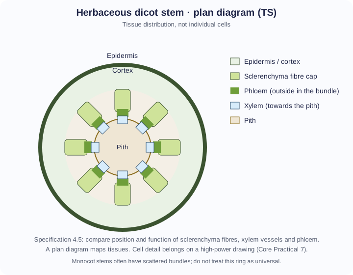
A plan diagramA drawing that shows the outlines of tissues and where they are, with no individual cells drawn. uses clear, single lines to show the outline of tissues: epidermis, cortex, vascular tissue, pith or mesophyll as appropriate. Do not sketch every cell, do not shade, and do not invent tissues you cannot see. Label xylemPlant tissue that carries water and mineral ions up from the roots. Its vessels are dead, hollow and lignified., phloemPlant tissue that transports sucrose and other organic substances, in sieve tubes with companion cells. and sclerenchymaPlant tissue of dead cells with thick lignified walls, such as fibres, that gives support. where their position is identifiable. Add a title, the organ, TS, and the magnificationHow many times larger an image is than the real object: magnification = image size ÷ actual size. or a scale from a calibrated graticule.
A high-power drawing of a small group of cells should show wall thickness, empty lumens, sieve plates if visible, and relative cell size. Draw only what is in the field. If a stain colours ligninA tough, waterproof polymer laid down in the walls of xylem vessels and sclerenchyma fibres., use that as evidence, but still describe the structure.
Identify from evidence
Xylem vessels: wide, thick lignifiedDescribes a cell wall strengthened and waterproofed by lignin. walls, empty-looking lumens, often towards the inside of a stem bundle or on the upper side of a leaf veinA vessel that carries blood back to the heart at low pressure. It has a thin wall, a wide lumen and valves.. Phloem: thin-walled living sieve tubeA phloem tube of living cells with few organelles, joined end to end through sieve plates. with small, densely stained companion cellA phloem cell next to a sieve tube element. It has many mitochondria and supports the sieve tube. beside them. Its position depends on the organ: outside the xylem in a stem bundle, below the xylem in a leaf vein, and between the arms of the xylem in a root. Sclerenchyma fibres: very thick lignified walls and tiny lumens (long cells, so in a TS you see them end-on as small, closely packed polygons), often as a cap outside the stem phloem or below a leaf vein.
In a typical dicot root, xylem may form a central star with phloem between the arms — do not copy the stem ring onto a root. If the section is oblique, shapes distort; say so. Repeat on more than one slide if available.
Variables, risk and evaluation
The organ is the independent comparison (root vs stem vs leaf). Dependent records are the labelled diagrams and identifications. Standardise microscope setup, drawing scale and the speciesA group of organisms that can interbreed to produce fertile offspring. if you are comparing organs from one plant. Staining, section thickness and how central the cut is all affect what you see.
Risks include broken slides and, if you cut your own sections, blades. Carry slides horizontally, cut away from fingers, and put damaged glass in the designated container. Do not use direct sunlight with a mirror illuminator.
The plant tissues microscope lab
Focus on stained sections of a stem, a root and a leaf, build a labelled plan, identify xylem vessels, sieve tubes and sclerenchyma fibres by position and structure, and measure a vessel with the graticule. The sections are procedurally drawn and every size is fictional.
Simulation only. Carry microscopes with two hands and slides by the edges, put broken glass in the glass bin, and never use the coarse focus at high power.
Method 7 steps
- Take prepared transverse sections (TS) of a stem, a root and a leaf. Carry slides flat and by the edges.
- Place the slide on the stage. Start with the ×4 objective and use the coarse focus (watching from the side as you bring the stage up) until the section is sharp.
- Centre the section, then draw a plan diagram: single clear lines around each tissue, no cells, no shading. Label the tissues; add a title, the organ, “TS” and the magnification or a scale.
- Centre one vascular bundle, change to ×10 and then ×40, refocusing with the fine focus only.
- Identify xylem vessels, phloem sieve tubes (with companion cells) and sclerenchyma fibres from their position and structure. Make a high-power drawing of a few cells of each.
- Measure structures with an eyepiece graticule calibrated for each objective, and use the measurement to add a scale.
- Repeat with the root TS and the leaf TS, and compare where each tissue is found.
Aligned with the order of the specification practical. Simulation only: a teacher sets the real volumes, concentrations and risk controls; this is not a laboratory protocol.
Variables
- Independent
- The organ (stem, root or leaf) whose section you examine.
- Dependent
- The tissues you identify and where they are: your labelled plan and high-power drawings, and your measurements.
- Controlled
- Same microscope, calibrated graticule and objective for comparable measurements
- Same stain and the same species of plant when comparing organs
- Thin, central (not oblique) sections
- Focus and illumination adjusted so walls are sharp without glare
- Same drawing rules for every plan
Safety
- Carry microscopes with two hands; carry slides flat and by the edges.
- Put broken glass in the designated container, not the bin, and tell the teacher.
- Never use the coarse focus at high power: the lens can hit and break the slide.
- If cutting your own sections, cut away from your fingers on a tile, and follow the school’s rules for stains.
- Never point a mirror illuminator at direct sunlight.
- Simulation only: the sections are procedural drawings and every size is a fictional teaching value.
Follow your school’s risk assessment; these notes are not a substitute for it.
Interpreting
- Stem (dicot): a ring of bundles; in each, sclerenchyma cap outermost, then phloem, cambium and xylem facing the pith.
- Root (dicot): a central xylem star with phloem between the arms, inside the endodermis; no pith.
- Leaf: in the midrib vein the xylem is on the upper (adaxial) side, the phloem below.
- Lignified walls (xylem and fibres) stain blue-green with toluidine blue; identify each tissue by position plus structure, not stain alone.
- This model shows dicots only. Monocot stems have scattered bundles, and the number of xylem arms in a root varies between species (2–6).
Guided method · step 1 of 11
In Slides under the simulator, drag Stem TS onto the stage, or press it.
Specification 4.5 locates the three named tissues in the stem, so it is the standard organ to start with.
1 · Plan and identify
Stem plan: 7 labels.
Slides
Objective and focus
Drag the stem TS onto the stage to begin.
2 · Measure a vessel
Calibration from Core Practical 5: ×4 = 25 µm, ×10 = 10 µm, ×40 = 2.5 µm per division. Measure the lumen (the clear inside), not the whole cell.
3 · Records and comparison
| Organ | Xylem | Phloem | Support |
|---|---|---|---|
| Stem TS | … | … | … |
| Root TS | … | … | … |
| Leaf TS | … | … | … |
Your cell identifications appear here.
Evaluate
Dicots only: monocot stems have scattered bundles and the number of root xylem arms varies between species. Light microscopes invert the image; this view moves with your drag for ease of use.
Make your reasoning visible
Plans have no cells
If you can count individual cells, you have drifted into a high-power drawing.
Label with evidence
“Thick-walled empty tube towards the pith” supports xylemPlant tissue that carries water and mineral ions up from the roots. Its vessels are dead, hollow and lignified. better than writing the word with no structure.
Avoid this misconception
The claim: Every circular plant section is a stem with a ring of bundles.
The correction: Roots and some stems have different layouts. Identify the organ first, then the tissues.
Check your understanding
Try each question before opening the suggested answer.
Practice question
Retrieval and explanation · suggested answer
State three rules of a plan diagram of a stem TS.
Show suggested answer
Show tissue outlines only, no individual cells or shading; label identifiable tissues; include title, TS and magnification or scale.
Practice question
Retrieval and explanation · suggested answer
How would you distinguish xylem vessels from sclerenchyma fibres at high power?
Show suggested answer
Both have lignified walls and can stain the same colour, so use structure and position. Xylem vessels have a wide, empty lumen inside the lignified wall, and sit on the inner (pith) side of a stem bundle. Sclerenchyma fibres are narrow, with a wall so thick the lumen is tiny, and are packed in a cap outside the phloem.
Practice question
Retrieval and explanation · suggested answer
Why might a leaf TS show xylem above phloem when a stem TS shows xylem towards the pith?
Show suggested answer
The organs have different polarities. In a typical dicot leaf vein, xylem is adaxial (upper); in a typical dicot stem bundle, xylem faces the centre. Always name the organ.
Take the idea further
Apply the same specification knowledge to an unfamiliar problem. Any extra assumptions are supplied in the question.
Stretch question
Your stem plan labels a dark-stained cap as xylem because “it is strong tissue”. Explain the error and how to correct the label.
Show a hint
Position plus cell type, not stain darkness alone.
Explore the reasoning
Sclerenchyma fibre caps also stain and are strong, but they sit outside the phloem. Xylem is the inner vessel tissue of the bundle. Correct the label using position (outside vs towards the pith) and cell shape (fibre vs wide vessel).
Recall this lesson
Lesson recall questions
Question 1 of 5: Does phloem transport mineral ions from root to leaf as its main job?
Xylem, phloem and sclerenchyma
Practice question
Retrieval and explanation · suggested answer
Does phloem transport mineral ions from root to leaf as its main job?
Try this from memory first.
Beyond the classroomFrom a school TS to a fibre cropCareers · plant anatomy · Enrichment, not required recall
Identifying flax or other fibre plants still starts with where the fibres sit relative to xylemPlant tissue that carries water and mineral ions up from the roots. Its vessels are dead, hollow and lignified. and phloemPlant tissue that transports sucrose and other organic substances, in sieve tubes with companion cells.. Kew’s speciesA group of organisms that can interbreed to produce fertile offspring. pages record fibre and other uses; they do not replace your own labelled drawing.
Back to the spec: 4.5–4.6: location of sclerenchyma fibres is part of both the specification and later materials use.
Kew POWO: Linum usitatissimumQuestion
Why would a plan of the wrong organ mislead a fibre harvest?
Explore a possible response
Fibre position differs between stem, root and leaf. Harvesting the wrong tissue wastes material and can destroy the vascular tissues the plant needs if it is still growing.