
What you need to know
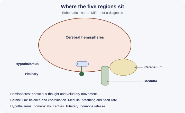
- 8.14Location and main functions of the cerebral hemispheres, hypothalamusA region of the brain that controls homeostasis, including body temperature and water balance., pituitary, cerebellum and medulla oblongata.
- 8.15MRI, fMRI, PET and CT are used to investigate brain structure and function.
Scans do not read thoughts. They are not a diagnosis you make from a lesson diagram. fMRI is function during a task. MRI structure is not the same image. No radiation doses to memorise.
By the end, you should be able to…
- State one main function for each of the five regions.
- Separate structure scans from function scans.
- Say which methods use ionising radiation.
Build your understanding
Five regions, five jobs
The cerebral hemispheres are the folded upper part of the brain. They deal with voluntary movement, interpretation of senses, language, memory and conscious thought. Different jobs sit in different areas. You do not need a phrenology map. “The thinking and voluntary part” is the right level, with one example if the question allows space.
Five regions, five jobs (continued)
The cerebellum sits at the back, under the hemispheres. It coordinates balance, posture and practised movement. It does not decide the goal of the movement. The medulla oblongata is the lowest part of the brainstem, continuous with the spinal cord. It contains centres for breathing and heart rateThe number of heartbeats per minute.. Damage there is immediately dangerous. The hypothalamusA region of the brain that controls homeostasis, including body temperature and water balance. is a small region under the hemispheres and above the pituitary. It contains homeostatic centres, including temperature and water balance, and it links the nervous system to the pituitary. The pituitary gland releases hormones that control other glands and some direct effects such as water balance. It hangs below the hypothalamus. Do not swap hypothalamus and pituitary: one is nervous tissue that controls, the other is the gland that releases the hormones.
Structure or activity
CT uses X-rays to make slices. The NHS describes it as detailed pictures with a small radiation risk if many scans are done. It shows structure. Dense material such as bone is clear. It is quick. It is not a map of which neurones were busy during a poem.
Structure or activity (continued)
MRI uses strong magnets and radio waves, not X-rays. NIBIB describes detailed pictures of soft tissues such as the brain, without ionising radiation. That is structure, and it is often better than CT for soft tissue. fMRI is the functional form NIBIB describes: it follows brain activation during a task, using changes linked to blood flow. A person might tap a finger or look at a picture. The coloured overlay is activity, not a photograph of thoughts. PET uses an injected radioactive tracer. The NHS describes that tracer and a small radiation exposure, and says PET can be combined with CT. Active tissue takes up more tracer, so PET is a functional image. It is not the same measurement as fMRI, and it does involve ionising radiation.
What a picture cannot say
A scan can show a region that is damaged or that is working during a task. It cannot, in this course, name a personality or prove what someone intended. Comparing the four methods means: ionising or not, structure or function, and what the person has to do during the scan.
The medulla’s control of breathing and heart rate is the same organ you may meet when discussing exercise. The specification here is the location and the main function, not a recalculation of cardiac outputThe volume of blood pumped by one ventricle per minute: cardiac output = stroke volume × heart rate..
| Method | What it uses | Structure or function |
|---|---|---|
| CT | X-rays | Structure |
| MRI | Magnets and radio waves | Structure, strong for soft tissue |
| fMRI | MRI during a task | Function, from blood-flow change |
| PET | Radioactive tracer | Function, where the tracer is used |
Make your reasoning visible
Gland versus centre
hypothalamusA region of the brain that controls homeostasis, including body temperature and water balance. detects and controls. Pituitary releases hormones.
f is the task
MRI is the anatomy. fMRI is the anatomy plus what is active during a task. PET is the tracer method.
Avoid this misconception
The claim: fMRI uses X-rays to read thoughts, and the cerebellum is the conscious part of the brain.
The correction: fMRI does not use X-rays and does not read thoughts. It maps activation during a task. Conscious thought is a cerebral job. The cerebellum coordinates movement.
Check your understanding
Try each question before opening the suggested answer.
Check your understanding (continued)
Practice question
Retrieval and explanation · suggested answer
State the main function of the cerebellum and of the medulla oblongata.
Show suggested answer
The cerebellum coordinates balance and movement. The medulla oblongata controls breathing and heart rate.
Practice question
Retrieval and explanation · suggested answer
How do the roles of the hypothalamus and the pituitary differ?
Show suggested answer
The hypothalamus contains homeostatic centres and controls the pituitary. The pituitary releases hormones.
Practice question
Retrieval and explanation · suggested answer
Which two of CT, MRI, fMRI and PET involve ionising radiation?
Show suggested answer
CT and PET. MRI and fMRI do not.
Practice question
Retrieval and explanation · suggested answer
What extra information does fMRI give compared with MRI?
Show suggested answer
Which regions are more active during a task, not only the shape of the tissue.
Take the idea further
Apply the same specification knowledge to an unfamiliar problem. Any extra assumptions are supplied in the question.
Stretch question
A student needs to know whether a soft-tissue region is missing, and separately whether that region is active while the person looks at pictures. No radioactive injection is allowed. Which methods fit the two questions, and which named method is ruled out by the injection limit?
Show a hint
Structure, then a task. Then look at PET.
Explore the reasoning
MRI answers the structure question for soft tissue without ionising radiation. fMRI answers the activity-during-looking question, also without a radioactive injection. PET is ruled out because it needs a radioactive tracer. CT could show structure but uses X-rays and is weaker for soft tissue, and it was not required once MRI is allowed.
Recall this lesson
Lesson recall questions
Question 1 of 5: State a main function of the medulla oblongata.
Main brain regions
Practice question
Retrieval and explanation · suggested answer
State a main function of the medulla oblongata.
Lesson: 8B 3: Investigating the Human Brain
Try this from memory first.
Put your biology to work
Explore where these ideas appear in the world. Enrichment, not extra required recall.
Activation during a task is not a thought transcript
8B · Investigating the Human BrainNIBIB describes MRI as magnet-and-radio pictures of soft tissue, and fMRI as a specialised MRI used to observe brain activation during cognitive tasks. That is the source for…Specification 8.15Read & use the scienceClose article
Why this matters
NIBIB describes MRI as magnet-and-radio pictures of soft tissue, and fMRI as a specialised MRI used to observe brain activation during cognitive tasks. That is the source for 8.15’s functional MRI. It does not say the scanner reads intentions. CT and PET are separate NHS descriptions: X-rays, and a radioactive tracer. 8.14 and 8.15: know the five regions, and know what each scan can show. A scan or growth response is indirect evidence: it cannot alone identify every signalling step or prove the proposed cause.
Back to the specification
Pearson Edexcel IAL Biology · Issue 2, February 2021 · Unit 5, pp. 35–38. The application extends beyond required recall.
- 8.15 MRI, fMRI, PET and CT in brain investigation.
Use the science
Why is “the fMRI showed the hypothalamus, so the person was thirsty” too strong?
Explore a possible response
Seeing a region, or even activation, is not a labelled thirst. Thirst would be an interpretation that needs the task and other evidence. The hypothalamus does contain water-balance centres, but a scan caption is not that conclusion by itself.
Explore the source
- NIBIB: magnetic resonance imagingUS National Institute of Biomedical Imaging and Bioengineering
PET traces molecules, not thoughts
8B · Investigating the Human BrainNIBIB explains that PET imaging uses a radioactive tracer whose distribution can be detected to investigate function in a living body. That is a different measurement from the…Specification 8.15Read & use the scienceClose article
Why this matters
NIBIB explains that PET imaging uses a radioactive tracer whose distribution can be detected to investigate function in a living body. That is a different measurement from the structural x-ray slices of CT or the magnetic signals of MRI. For 8.15, the distinction matters more than the colours on a scan: each technique turns a physical signal into an image with limits and uncertainty. A bright PET region is not a transcript of a person’s thoughts. Researchers or clinicians must know which tracer was used, when the measurement was made and what comparison is appropriate before interpreting a pattern.
Back to the specification
Pearson Edexcel IAL Biology · Issue 2, February 2021 · Unit 5, pp. 35–38. The application extends beyond required recall.
- 8.15 MRI, fMRI, PET and CT in brain investigation.
Use the science
Why might a PET image and an MRI image of the same brain answer different biological questions?
Explore a possible response
PET maps the distribution of a selected tracer and can reflect a function or biochemical process. MRI provides detailed structure; the signals and interpretations are different.
Explore the source
- Nuclear medicine and PETUS National Institute of Biomedical Imaging and Bioengineering