
What you need to know
- 3.1Cells share common features.
- 3.3(i–ii)eukaryoticDescribes cells with a nucleus and membrane-bound organelles, such as animal and plant cells. organelleA structure inside a cell with a particular function, such as a mitochondrion or ribosome. ultrastructureThe detailed structure of a cell as seen with an electron microscope. and functions.
- 3.6Recognition of organelles in electron micrographs.
By the end, you should be able to…
- Link each specified organelle to its function.
- Use structural clues rather than colour to identify organelles.
- Explain why a single section may omit a structure.
Build your understanding
A shared cellular plan with compartments
Cells have a cell surface membraneThe partially permeable membrane around a cell that controls which substances enter and leave. enclosing cytoplasmEverything inside the cell surface membrane except the nucleus: the fluid cytosol and the organelles.. Living cells share the molecular machinery of life, including genetic information and ribosomeA small organelle made of rRNA and protein, where translation takes place., although specialised mature cells may lose some structures. eukaryoticDescribes cells with a nucleus and membrane-bound organelles, such as animal and plant cells. cells characteristically compartmentalise processes in membrane-bound organelleA structure inside a cell with a particular function, such as a mitochondrion or ribosome..
A typical eukaryotic cell contains a nucleusThe organelle that contains the cell’s chromosomes, surrounded by a nuclear envelope with pores., but do not turn a typical plan into an absolute claim: mature mammalian red blood cells, for example, lack nuclei. The particular organelles and their abundance reflect cell function.
Nucleus, nucleolus and ribosomes
The nucleus has a double-membrane envelopeAn outer covering membrane. The nucleus and chloroplasts have a double-membrane envelope; some viruses, such as HIV, have a lipid envelope taken from the host cell. with pores controlling exchange with the cytoplasm. It contains chromatin, including DNADeoxyribonucleic acid: a double-stranded polynucleotide that carries the genetic code., and is the site of transcriptionMaking an mRNA copy of a gene, using one strand of DNA as a template, in the nucleus. in the eukaryotic model. The nucleolusA dense region in the nucleus where rRNA is made and ribosomes are assembled. is a dense region, not a separate membrane-bound organelle; it produces rRNARibosomal RNA: RNA that, with proteins, makes up a ribosome. and assembles ribosomal subunits.
Ribosomes are small, non-membrane-bound structures made of rRNA and protein. They synthesise polypeptides. Cytoplasmic eukaryotic ribosomes are conventionally described as 80S; they may be free or attached to rough ER. Free ribosomes make many proteins used within the cytosol, while bound ribosomes make proteins entering the secretory pathway.
Membranes for manufacture and processing
rough endoplasmic reticulumA network of flattened membrane sacs covered in ribosomes. It makes and transports proteins. consists of flattened membrane sacs with ribosomes on the cytosolic surface. It supports synthesis, folding and transport of proteins entering its lumenThe space inside a tube, such as a blood vessel. or membrane. Smooth ER lacks attached ribosomes and has roles in lipid synthesis; some cells also use it in detoxification or calcium storage.
The Golgi apparatusA stack of flattened membrane sacs that modifies proteins and packages them into vesicles. is a stack of flattened membrane sacs with associated vesicles. It modifies, sorts and packages proteins and lipids received from the ER. lysosomeA vesicle containing hydrolytic (digestive) enzymes. are membrane-bound compartments containing hydrolytic enzymeA globular protein that acts as a biological catalyst, speeding up a specific reaction. for intracellular digestion and recycling. Their contents and appearance vary, so a dark round body alone is not conclusive identification.
Mitochondria and centrioles
mitochondrionThe organelle where the link reaction, Krebs cycle and oxidative phosphorylation of aerobic respiration take place. have an outer membrane and a folded inner membrane forming cristaeThe folds of the inner mitochondrial membrane. They give a large surface area for the electron transport chain and ATP synthase. around a matrix. They carry out aerobic respirationRespiration using oxygen: glycolysis, the link reaction, the Krebs cycle and oxidative phosphorylation. It releases much more ATP than anaerobic respiration. and much of the cell’s ATPAdenosine triphosphate: the molecule that releases energy for cell processes when it is hydrolysed to ADP and Pi. production. The folds provide a large membrane area for reactions involved in ATP synthesis. Detailed respirationThe breakdown of respiratory substrates such as glucose in cells to release energy, which is used to make ATP. pathways are developed later in the course.
Centrioles are cylindrical arrangements of microtubules, commonly found as a pair at right angles in an animal-cell centrosome. A cross-section may show nine triplets of microtubules. They are associated with microtubule organisation and spindle formation; do not confuse them with chromosomeA long DNA molecule wound around histone proteins. After replication it is two sister chromatids joined at a centromere. centromeres.
Read the image, not a memorised cartoon
Use several clues together: membranes, internal folds, ribosome dots, stacked sacs, position and scale. A mitochondrial section might be circular or elongated depending on the cutting plane. A nucleus can be absent from a particular thin section even though the complete cell contains one.
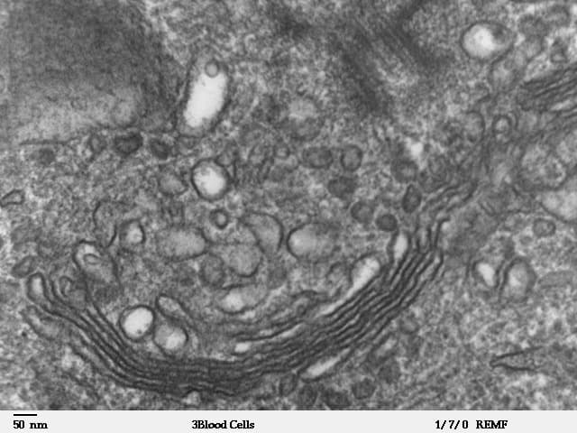
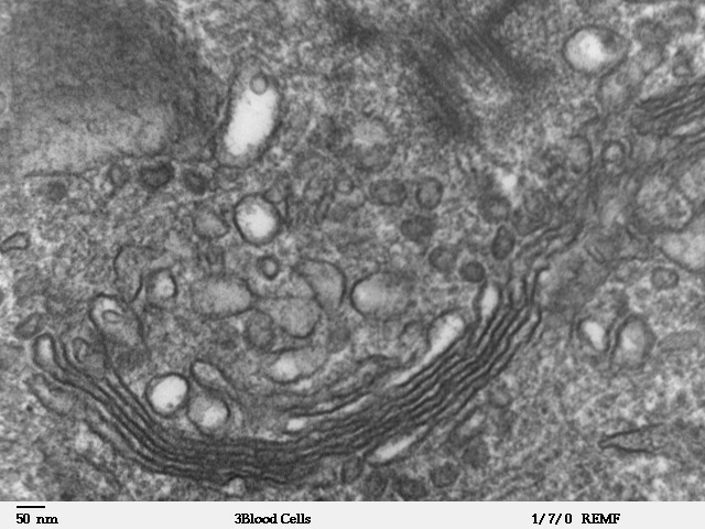
In the micrograph below, look for the curved stack of flattened membranes across the lower middle and the many nearby rounded membrane profiles. These are evidence for a Golgi region with associated vesicles. The image is a real TEM of part of a human leukocyte, not a complete cell diagram. Do not identify every dark spot as a ribosome or claim to see all the organelles in this one image.
NOT: the nucleolus is not a membrane-bound organelle, and a centriole is not a centromere.
- Rough ER has ribosomes; smooth ER does not
- Mitochondrial cristae increase membrane area
- Lysosome contents vary, so a dark circle is weak evidence alone
- Mature red blood cells lack nuclei
Two traps this diagram blocks
- Do not identify an organelle by the colour added to a micrograph.
- Do not say a structure is absent from the cell because it is missing from one thin section.
From the diagram alone
Question
Describe how the structure of a mitochondrion is related to its function. [3]
Show a model answer
It has a double membrane. The inner membrane is folded into cristae, which gives a large area for the reactions that produce ATP in aerobic respiration. The matrix is the compartment inside those folds.
| Structure | Typical EM clue | Main function |
|---|---|---|
| Nucleus | Envelope, pores, chromatin | Contains DNA; transcription |
| Nucleolus | Dense region within nucleus | rRNA and ribosomal subunit assembly |
| Ribosomes | Small dense particles, free or on rER | Polypeptide synthesis |
| rER | Membrane sacs with surface ribosomes | Protein synthesis and transport |
| sER | Tubular membranes without ribosomes | Lipid synthesis |
| Golgi | Stacked sacs with vesicles | Modification, sorting and packaging |
| Mitochondrion | Double membrane and cristae | Aerobic ATP production |
| Lysosome | Membrane-bound body, variable dense contents | Intracellular digestion |
| Centriole | Microtubule cylinder; triplets in cross-section | Microtubule organisation |
Make your reasoning visible
Give structure and consequence
For mitochondrionThe organelle where the link reaction, Krebs cycle and oxidative phosphorylation of aerobic respiration take place., link inner-membrane folds to membrane area and ATPAdenosine triphosphate: the molecule that releases energy for cell processes when it is hydrolysed to ADP and Pi. production. “It has cristaeThe folds of the inner mitochondrial membrane. They give a large surface area for the electron transport chain and ATP synthase.” alone is only a description.
Name the synthesis site correctly
ribosomeA small organelle made of rRNA and protein, where translation takes place. synthesise polypeptides; the Golgi modifies and sorts them. The nucleolusA dense region in the nucleus where rRNA is made and ribosomes are assembled. assembles ribosomal subunits.
Use multiple image clues
A shape alone is weak evidence. Combine membranes, internal features and context, and state uncertainty where needed.
Key explanations
- The nucleusThe organelle that contains the cell’s chromosomes, surrounded by a nuclear envelope with pores. is bounded by an envelopeAn outer covering membrane. The nucleus and chloroplasts have a double-membrane envelope; some viruses, such as HIV, have a lipid envelope taken from the host cell. with pores and contains chromatin. transcriptionMaking an mRNA copy of a gene, using one strand of DNA as a template, in the nucleus. occurs there in a eukaryoticDescribes cells with a nucleus and membrane-bound organelles, such as animal and plant cells. cell.
Avoid this misconception
The claim: Every organelleA structure inside a cell with a particular function, such as a mitochondrion or ribosome. is surrounded by a membrane.
The correction: Ribosomes and centrioles are not membrane-bound, and the nucleolus is a region inside the nucleus.
What do animal, plant and bacterial cells share, and what sets them apart?
The same structure names are used for all three. Presence and absence tell them apart, not inventing new names for the same part.
What the model shows
An animal cell, a plant cell and a bacterium with the same label list, a detail for each structure and a comparison table.
What to look for
Which structures all three have. Which are eukaryotic only. How plant and bacterial walls differ.
What it tells us
Shared features are a membrane, cytoplasm, DNA and ribosomes. Membrane-bound organelles mark the eukaryotic plan. Walls differ in cellulose versus peptidoglycan.
Be careful what you claim
The drawings are teaching diagrams, not micrographs, and the bacterium is not to scale.
Try it in the model: select Ribosomes on the animal cell, then switch to Bacterium and select Ribosomes again. Compare the detail text.
Three cells, the same labels
Choose an animal cell, a plant cell or a bacterium, then select a structure. The table uses the same list for all three. Colour only separates the parts. This is a teaching diagram, not a micrograph, and the bacterium is drawn large enough to label.
What you see
- A labelled diagram of an animal cell, a plant cell or a bacterium. Colour only separates the parts.
- A detail panel for the structure you select, saying whether it is present, in some cells, or absent.
- A table of the same structures across all three cell kinds.
How to use it
- Choose the cell kind, then click a structure on the diagram or in the list.
- Use Hide wall and capsule to see inside a plant cell or a bacterium more clearly.
What it shows
- Shared features are a cell-surface membrane, cytoplasm, DNA and ribosomes (3.3, 3.5).
- Eukaryotic cells have a membrane-bound nucleus and membrane-bound organelles; a bacterium has a nucleoid and none of those organelles.
- A plant wall is cellulose; a bacterial wall contains peptidoglycan. Capsules, plasmids, pili and flagella are not in every bacterium.
Limits of the model
- This is a teaching diagram, not a micrograph. The bacterium is drawn large enough to label; real bacterial cells are usually much smaller.
- A typical plan is shown. Mature mammalian red blood cells are eukaryotic exceptions with no nucleus.
- Plant chloroplasts and the large vacuole are named here and studied further in Topic 4.
Guided tour · step 1 of 8
- In Structures, just below these steps, keep Cell on Animal cell (options: Animal cell, Plant cell, Bacterium).
- Press the Nucleus button, or click the nucleus in the diagram.
- Read the detail and the table row.
A typical animal cell is eukaryotic: a membrane-bound nucleus and membrane-bound organelles.
Structures
Questions
Comparison table
| Structure | Animal | Plant | Bacterium |
|---|---|---|---|
| Cell-surface membrane | Present | Present | Present |
| Cytoplasm | Present | Present | Present |
| Ribosomes | Present | Present | Present |
| Nucleus | Present | Present | Absent |
| Nucleolus | Present | Present | Absent |
| Rough ER | Present | Present | Absent |
| Smooth ER | Present | Present | Absent |
| Golgi apparatus | Present | Present | Absent |
| Vesicle | Present | Present | Absent |
| Mitochondrion | Present | Present | Absent |
| Lysosome | Present | Present | Absent |
| Centrioles | Present | Absent | Absent |
| Cell wall | Absent | Present | Present |
| Chloroplast | Absent | Present | Absent |
| Large permanent vacuole | Absent | Present | Absent |
| Nucleoid and circular chromosome | Absent | Absent | Present |
| Plasmid | Absent | Absent | Some cells |
| Capsule | Absent | Absent | Some cells |
| Pili | Absent | Absent | Some cells |
| Flagellum | Absent | Absent | Some cells |
Shared features are a membrane, cytoplasm, DNA and ribosomes. The eukaryotic differences to name are a membrane-bound nucleus and membrane-bound organelles. A plant wall is cellulose. A bacterial wall contains peptidoglycan. Capsules, plasmids, pili and flagella are not in every bacterium. Centrioles belong to the animal plan used here, not the plant cell.
Check your understanding
Try each question before opening the suggested answer.
Practice question
Retrieval and explanation · suggested answer
An EM profile has a double membrane and internal folds. Suggest an organelle and explain the advantage of the folds.
Show suggested answer
A mitochondrion. Cristae increase inner-membrane surface area for reactions involved in ATP production.
Practice question
Retrieval and explanation · suggested answer
Why would a cell exporting large quantities of protein often have extensive rough ER and Golgi?
Show suggested answer
Bound ribosomes and rough ER support synthesis and processing; the Golgi modifies and packages the proteins for transport.
Practice question
Retrieval and explanation · suggested answer
In the supplied TEM, which visible features support identifying the Golgi region?
Show suggested answer
A curved stack of flattened membrane sacs in the lower central region, with nearby rounded vesicle profiles. This combination is stronger evidence than a single dark line.
Take the idea further
Apply the same specification knowledge to an unfamiliar problem. Any extra assumptions are supplied in the question.
Stretch question
A thin section contains no visible nucleus. Can you classify the cell as prokaryotic from that observation alone?
Show a hint
Think about what a thin slice samples.
Explore the reasoning
No. The section may pass through cytoplasm away from a eukaryotic nucleus. Look for other evidence such as membrane-bound organelles, additional sections and the scale of structures.
Recall this lesson
Lesson recall questions
Question 1 of 5: How does a plasma membrane count as a common cellular feature across organisms?
Cells as a shared organisation
Practice question
Retrieval and explanation · suggested answer
How does a plasma membrane count as a common cellular feature across organisms?
Lesson: 3A 4: Prokaryotic Cells
Try this from memory first.
Put your biology to work
Explore where these ideas appear in the world. Enrichment, not extra required recall.
Interpreting a real cell section
3A · Eukaryotic Cells 1: Common Cellular StructuresLouisa Howard’s TEM image shows a Golgi region in a human leukocyte. Researchers interpret features in prepared sections rather than expecting every structure to resemble a textbook drawing.…Specification 3.6Read & use the scienceClose article
Why this matters
Louisa Howard’s TEM image shows a Golgi region in a human leukocyte. Researchers interpret features in prepared sections rather than expecting every structure to resemble a textbook drawing. A thin section shows only one slice of a three-dimensional cell. An apparent isolated circle may be part of a larger membrane system outside the plane of section. The Golgi can be identified more confidently when several flattened sacs and nearby vesicles are seen together. A clinical scientist examining a leukocyte would describe the visible evidence before naming a structure. Preparation and staining also create contrast and sometimes artefacts. The exam skill is to identify features from evidence, not from a perfect diagram.
Back to the specification
Pearson Edexcel IAL Biology · Issue 2, February 2021 · Unit 2, pp. 21–24. The application extends beyond required recall.
- 3.6 be able to recognise the organelles in 3.3 from electron microscope (EM) images
Use the science
What additional evidence would help identify an ambiguous small membrane-bound profile?
Explore a possible response
Other sections, higher-resolution detail or specific labelling could help; shape alone may fit several structures.
Explore the source
- Louisa Howard / Dartmouth: Golgi TEM, public domainWikimedia Commons

The stain that makes a diagnosis possible
3A · Eukaryotic Cells 1: Common Cellular StructuresPathologists examine thin, stained tissue sections to identify disease. The US National Cancer Institute explains that routine dyes make nuclei, cytoplasm and tissue organisation easier to distinguish. Staining…Specification 3.7(ii) · 3.3(i)Read & use the scienceClose article
Why this matters
Pathologists examine thin, stained tissue sections to identify disease. The US National Cancer Institute explains that routine dyes make nuclei, cytoplasm and tissue organisation easier to distinguish. Staining adds contrast; it does not manufacture an organelle that was absent before preparation. A cell in a section may look different from a complete textbook drawing because the cut passes through only part of it. A pathologist also considers the surrounding tissue and may use molecular tests. This career example turns the specification’s microscopy and organelle recognition into a practical evidence question: which visible feature supports the proposed identification, and what remains uncertain?
Back to the specification
Pearson Edexcel IAL Biology · Issue 2, February 2021 · Unit 2, pp. 21–24. The application extends beyond required recall.
- 3.7(ii) understand the importance of staining specimens in microscopy
- 3.3(i) know the ultrastructure of eukaryotic cells, including nucleus, nucleolus, ribosomes, rough and smooth endoplasmic reticulum, mitochondria, centrioles, lysosomes and Golgi apparatus
Use the science
A dark patch appears after staining a tissue section. Why is it unsafe to call it a nucleus from colour alone?
Explore a possible response
Dyes bind several structures and the section is only one plane. Position, shape, surrounding features and possibly a second stain or molecular test are needed.
Explore the source
- Pathology reports and stained tissue sectionsNational Cancer Institute