
What you need to know
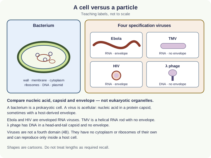
- 6.5(i)Viruses as acellular particles with nucleic acid in a capsidThe protein coat of a virus, made of capsomeres..
Historical method is enrichment. Required comparisons remain Ebola, TMV, HIV and λ phage.
By the end, you should be able to…
- Explain why a filterable, cell-free agent required a new category.
- Link TMVTobacco mosaic virus: a rod-shaped RNA virus that infects plants.’s RNARibonucleic acid: a single-stranded polynucleotide with ribose and uracil. mRNA, tRNA and rRNA take part in protein synthesis. rod to the 6.5(i) headings.
- Refuse extra plant-virus lists as automatic specification content.
Build your understanding
Smaller than a filter that stops bacteria
Nineteenth-century tobacco disease could be transferred by sap that had passed through filters that retained bacteria. The agent was reproducing in the plant, yet it was not a cell on a plate. That method story is why “virus” had to mean something other than “tiny bacterium”.
APSNet’s TMVTobacco mosaic virus: a rod-shaped RNA virus that infects plants. pages describe a rod of RNARibonucleic acid: a single-stranded polynucleotide with ribose and uracil. mRNA, tRNA and rRNA take part in protein synthesis. plus protein, about 300 nm long in their teaching figure — a size for context, not a number you must recall. School 6.5 still wants RNA, helical capsidThe protein coat of a virus, made of capsomeres., no envelopeAn outer covering membrane. The nucleus and chloroplasts have a double-membrane envelope; some viruses, such as HIV, have a lipid envelope taken from the host cell..
A particle that needs a host
TMV does not grow on nutrient agar. It has no cytoplasmEverything inside the cell surface membrane except the nucleus: the fluid cytosol and the organelles. to run a lag-exponential curve of its own. Measuring “growth” of a virus means counting particles or infected hosts, not CFU of the virus as a free-living colony. That is why 6.2–6.4 are written for microorganisms in culture, with viruses compared as structures in 6.5.
Do not turn the history into extra named plant viruses. EbolaAn enveloped RNA virus that causes Ebola virus disease, a severe haemorrhagic fever., HIVHuman immunodeficiency virus: an RNA retrovirus that infects T helper cells and can lead to AIDS. and lambda phageA bacteriophage that infects E. coli and can follow either a lytic or a lysogenic cycle. remain the other three specified comparisons.
Make your reasoning visible
Method, then category
Filterable and acellular, therefore not a bacterium — then apply nucleic acid, capsidThe protein coat of a virus, made of capsomeres., envelopeAn outer covering membrane. The nucleus and chloroplasts have a double-membrane envelope; some viruses, such as HIV, have a lipid envelope taken from the host cell..
Do not inflate the list
A research paper’s extra virus is not automatic 6.5 content.
Avoid this misconception
The claim: TMVTobacco mosaic virus: a rod-shaped RNA virus that infects plants. is a bacterium that lost its wall.
The correction: TMV is an RNARibonucleic acid: a single-stranded polynucleotide with ribose and uracil. mRNA, tRNA and rRNA take part in protein synthesis. virus with a helical capsid and no envelope. It never was a prokaryotic cell.
Check your understanding
Try each question before opening the suggested answer.
Practice question
Retrieval and explanation · suggested answer
Why did a bacteria-stopping filter still allow TMV to transmit disease?
Show suggested answer
The agent is a virus particle smaller than a bacterium, so it could pass the filter and still infect the plant.
Practice question
Retrieval and explanation · suggested answer
Which 6.5 headings still apply to TMV after this history?
Show suggested answer
RNA nucleic acid, helical capsid, no envelope; acellular, reproduces only in a host.
Take the idea further
Apply the same specification knowledge to an unfamiliar problem. Any extra assumptions are supplied in the question.
Stretch question
A student wants to plot a four-phase growth curve for TMV in a flask of broth. What category error is that, and what would you measure instead if you were studying spread in plants?
Show a hint
No cytoplasm, no binary fission in broth.
Explore the reasoning
TMV is not a free-living cell, so lag–exponential–stationary–death in broth does not apply. You would need a host (plants or protoplasts) and a measure of particles or lesions, which is not Core Practical 13.
Recall this lesson
Lesson recall questions
Question 1 of 5: Compare the nucleic acid of a bacterium with that of a virus.
Bacteria vs viruses
Practice question
Retrieval and explanation · suggested answer
Compare the nucleic acid of a bacterium with that of a virus.
Lesson: 6A 1: Bacteria and Viruses
Try this from memory first.
Put your biology to work
Explore where these ideas appear in the world. Enrichment, not extra required recall.
A teaching virus that earned its place
6A · Thinking BiggerAPSNet presents TMV as stable, well studied and useful for showing virus–host interaction. That is why it is the plant example in 6.5(i), not because tobacco farming is…Specification 6.5(i)Read & use the scienceClose article
Why this matters
APSNet presents TMV as stable, well studied and useful for showing virus–host interaction. That is why it is the plant example in 6.5(i), not because tobacco farming is on the specification. Treat their 300 nm rod as a published teaching size, not a recall fact. TMV was useful because its simple, robust particle could be purified and studied. A named virus still has a host and a genome type; historical fame does not replace those structural comparisons.
Back to the specification
Pearson Edexcel IAL Biology · Issue 2, February 2021 · Unit 4, pp. 29–32. The application extends beyond required recall.
- 6.5(i) be able to compare the structure of bacteria and viruses (nucleic acid, capsid structure and envelope) with reference to Ebola virus, tobacco mosaic virus (TMV), human immunodeficiency virus (HIV) and lambda phage (λ phage)
Use the science
Why is “TMV is 300 nm, therefore all viruses are 300 nm” wrong?
Explore a possible response
That length is one teaching measurement for TMV. Ebola, HIV and λ phage have different shapes and sizes; the required comparison is nucleic acid, capsid and envelope.
Explore the source
- APSNet: tobacco mosaic virus, purpose and backgroundAmerican Phytopathological Society
The Ebola image is a structure, not a monster
6A · Bacteria and VirusesFilm posters often turn a virus into a spiky creature with a face. A CDC electron micrograph instead shows a filamentous Ebola particle with an outer envelope and…Specification 6.5(i)Read & use the scienceClose article
Why this matters
Film posters often turn a virus into a spiky creature with a face. A CDC electron micrograph instead shows a filamentous Ebola particle with an outer envelope and internal nucleocapsid. The shape may help a researcher recognise a family, but the image cannot reveal the RNA sequence or prove how that particular particle will behave in a host. Ebola is one of four specified viruses to compare by nucleic acid, capsid and envelope. A bacterium has a cellular structure with cytoplasm and ribosomes; the virus does not. A dramatic image is a starting observation, not a complete biological classification.
Back to the specification
Pearson Edexcel IAL Biology · Issue 2, February 2021 · Unit 4, pp. 29–32. The application extends beyond required recall.
- 6.5(i) be able to compare the structure of bacteria and viruses (nucleic acid, capsid structure and envelope) with reference to Ebola virus, tobacco mosaic virus (TMV), human immunodeficiency virus (HIV) and lambda phage (λ phage)
Use the science
What can the electron image support about Ebola, and what would require a different test?
Explore a possible response
It can support observations about particle shape and envelope projections. Genome type or sequence needs molecular analysis, not shape alone.
Explore the source
- Ebola virus electron micrographCDC Public Health Image Library