
By the end of this lesson
- Identify the major structures in typical animal and plant cells.
- Interpret cell diagrams without assuming that shape, colour or position is fixed.
- Distinguish cell structures from cells and tissues.
BIG QUESTION
How can a flat diagram represent a living cell that is three-dimensional, dynamic and mostly colourless?
Cell diagrams simplify shape and colour so structures can be identified, but the simplification must not be mistaken for the living cell itself.
A cell diagram is a model
Living cells are three-dimensional and their organelles move within the cytoplasm. Textbook diagrams use clear outlines, artificial colours and separated labels to make structures visible. The colours do not usually show the natural colour of the structures.
A section through a cell may miss an organelle or cut it in an unfamiliar way. Identification should therefore use several clues: boundary, relative position, shape, number and the type of cell shown.
Structures shared by typical animal and plant cells
Both typical animal and plant cells contain a cell membrane, cytoplasm, nucleus, mitochondria and ribosomes. These are eukaryotic structures, although not every specialised cell retains every structure—for example, a mature mammalian red blood cell has no nucleus.
The cell membrane forms the cell boundary. Cytoplasm fills much of the interior. The nucleus is usually one of the largest organelles, mitochondria are smaller and numerous, and ribosomes are too small to be resolved individually with a school light microscope.
Structures characteristic of many plant cells
A typical plant cell also has a celluloseA polysaccharide made of glucose that forms plant cell walls; it is the main part of dietary fibre in human food. outside its membrane and a large permanent vacuoleA large space in a plant cell filled with cell sap; it keeps the cell turgid, which supports the plant. containing cell sap. Photosynthetic plant cells contain chloroplastAn organelle in plant cells that contains chlorophyll and is the site of photosynthesis..
Do not turn “typical” into “all.” Root hair cells are plant cells but usually lack chloroplasts because roots receive little light. Some plant cells lose their living contents at maturity, as happens in xylem vessels.
Reading the two boundaries of a plant cell
The plant cell wallA freely permeable layer outside the cell membrane that supports the cell; in plants it is made mainly of cellulose (fungal walls contain chitin). is the thicker outer boundary. The cell membraneA selectively (partially) permeable layer around a cell that controls which substances enter and leave. lies just inside it and controls exchange with the surroundings. They are separate structures with different properties.
The vacuole can occupy most of a mature plant cell, pushing the cytoplasm and nucleus towards the edge. A diagram with a large central space is therefore not showing an “empty” cell.
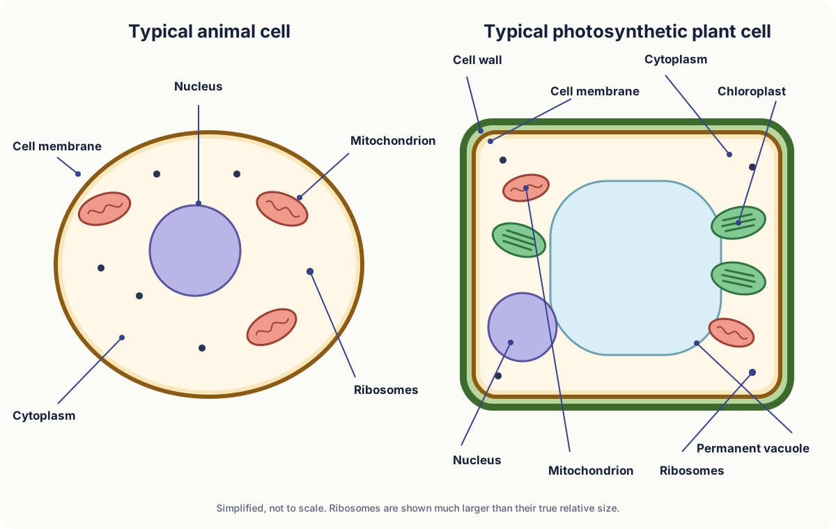
Use position and boundaries as identification clues
No single clue should be used in isolation.
| Structure | Useful visual clue | Caution |
|---|---|---|
| Cell membrane | Thin boundary around the cytoplasm. | In a plant cell it lies just inside the cell wall. |
| Nucleus | Often a large rounded organelle. | Position varies and some specialised cells lack one. |
| Mitochondrion | Small oval or bean-shaped organelle. | Usually many are present; not individually visible at low resolution. |
| Ribosome | Shown as tiny dots in diagrams. | Too small to resolve with a school light microscope. |
| Cell wall | Rigid outer boundary of a plant cell. | It is outside, and separate from, the membrane. |
| Permanent vacuole | Large fluid-filled compartment. | Its membrane is the tonoplast, usually beyond this specification. |
| Chloroplast | Oval organelle, often drawn green. | Found in photosynthetic cells, not every plant cell. |
Language that earns precision
- cell membrane
- The selectively permeable boundary surrounding the cell contents.
- cytoplasm
- The cell material where many enzyme-controlled reactions occur.
- nucleus
- An organelle containing most of a eukaryotic cell’s genetic material.
- mitochondrion
- An organelle where aerobic respiration releases energy.
- ribosome
- A small structure where proteins are synthesised.
- cell wall
- A freely permeable cellulose layer that supports a plant cell.
- permanent vacuole
- A membrane-bound compartment containing cell sap in a mature plant cell.
- chloroplast
- An organelle containing chlorophyll where photosynthesis occurs.
Common misconceptions
Claim: The cell wall replaces the cell membrane in plants.
Correction: Plant cells have both: the membrane controls exchange and the wall provides support.
Claim: All plant cells contain chloroplasts.
Correction: Only photosynthetic plant cells require chloroplasts; most root cells do not contain them.
Claim: Cells and organelles naturally have textbook colours.
Correction: Most are colourless; stains and false colour increase contrast.
Claim: The vacuole is an empty gap.
Correction: It is a membrane-bound compartment filled with cell sap.
Match the answer to the command word
Give the structure’s name; use boundaries and position together rather than colour alone.
Use a straight line that ends on the precise structure and does not cross another label line.
State visible features only; save functions for an explain question.
Recall, apply, then justify
Recall, apply, then justify (continued)
Name five structures found in both a typical animal cell and a typical plant cell.
Hint
Exclude the three structures usually shown as plant-only.
Suggested marking points
- Cell membrane.
- Cytoplasm.
- Nucleus.
- Mitochondria.
- Ribosomes.
A diagram of a root hair cell contains no chloroplasts. Explain why this can still be a plant cell.
Hint
Plant identity does not depend on one organelle; connect location to light.
Suggested marking points
- It has plant features such as a cellulose cell wall and permanent vacuole.
- Root cells are usually underground and do not receive enough light for photosynthesis, so chloroplasts are unnecessary.
Distinguish the cell wall from the cell membrane in a plant-cell diagram.
Hint
State both relative position and appearance.
Suggested marking points
- The cell wall is the thicker outer boundary.
- The cell membrane is the thin boundary immediately inside it.
Suggest why ribosomes are shown in a textbook diagram but are not visible in a school light-microscope image.
Hint
Compare the object size with resolving power.
Suggested marking points
- Ribosomes are smaller than the resolution limit of a light microscope.
- A diagram can represent structures that cannot be resolved in that image.
Evaluate the limitations of using one labelled two-dimensional cell diagram as evidence for the structure of all plant cells.
Planning hint
Balance what the model communicates clearly against variation and information it omits.
What a strong design includes
- It clearly communicates the main structures and their relative locations.
- A single section cannot show the whole three-dimensional cell.
- Plant cells differ by tissue and function, so not all contain chloroplasts or a large vacuole.
- Colours, sizes, organelle numbers and positions are simplified.
- A conclusion should state that the diagram is useful as a typical model but not a complete record of every plant cell.